
Background: The rapid integration of artificial intelligence (AI) and medical big data into health care is transforming diagnosis, treatment planning, and research. However, formal education in these areas remains limited in undergraduate medical curricula, particularly in China. Objective: This study aimed to investigate clinical medicine undergraduates’ familiarity with AI and medical big data, their perceived need for related courses, and their preferred curriculum design and assessment methods. Methods: A cross-sectional, web-based survey was conducted at Zunyi Medical University, Guizhou, China, from January 10 to 17, 2025. In the institutional context of this study, “clinical medicine” included related clinical-track specialties such as pediatrics and psychiatry. All eligible students (N=1094) were invited, and 871 (79.6%) were included in the final analysis. The self-administered questionnaire was developed based on a literature review and expert consultation, with content validity quantified using the content validity index. Descriptive statistics were used to summarize response distributions. For ordinal outcomes (items 1-14), adjusted ordinal logistic regression models were applied, with gender and grade as predictors and major as a covariate. Given the small number of third- and fourth-year students, grade was modeled as an ordered trend variable. For nominal outcomes (items 15-16), group differences were assessed using chi-square tests or Fisher exact tests, as appropriate. Results: A total of 871 students were analyzed, of whom 62.6% (n=545) were women. Overall familiarity with AI and medical big data was limited: 34.8% (303/871) agreed or strongly agreed that they were familiar with the topic, and only 33% (287/871) reported having at least some prior learning experience. In contrast, the perceived educational need was high: 94% (819/871) considered such a course at least somewhat necessary, 57% (497/871) reported that the course was needed or very needed, 75.5% (658/871) indicated that they would likely or definitely enroll, and 56.5% (492/871) reported that they would likely or definitely engage in self-directed learning. Personalized teaching based on textbooks (566/871, 65%) or open-book examinations (633/871, 72.7%) was the most preferred instructional and assessment format. Preferences for course materials and assessment methods differed by grade but not by gender. Conclusions: Early-stage clinical medicine undergraduates demonstrated limited familiarity with AI and medical big data but expressed a strong demand for related education. Students preferred structured yet flexible instructional formats and open-book assessments. Although the findings are based predominantly on first- and second-year students, they support the development of staged, practice-oriented AI and medical big data curricula tailored to the needs of early-stage clinical medicine undergraduates.
<img src="https://jmir-production.s3.us-east-2.amazonaws.com/thumbs/a22edb5d5bd9dfd0ef9d0e91d5560eaf" />
Why California’s carbon manure math doesn’t add up
Something stinks in California’s climate policies.
Years ago, the state set up a system that pays cattle farmers across the country to turn the methane emitted from cattle manure into natural gas, encouraging the dairy sector to produce a gas we burn instead of one that just pollutes the air.
It’s become wildly popular because the subsidies are extremely lucrative. But a growing body of research suggests the program is a case study in the shortcomings of our preferred approaches to climate action. Instead of simply forcing industries to directly cut their pollution or pay for it as a cost of doing business, legislators have repeatedly opted to set up convoluted incentive systems that swap climate responsibilities between parties and regions. As studies have shown again and again, these carbon offsetting and trading schemes often dramatically overstate the emissions reductions actually achieved in the one place that matters: the atmosphere.
The dairy program illustrates a particular version of this problem, muddling the impacts of different types of greenhouse gases in a way that researchers argue will lock in more warming in the future.
Despite this and other concerns, California regulators decided in 2024 to extend parts of the program beyond 2050. And a recent proposal by the state’s air resources board could send millions of additional dollars to dairy farmers as part of a plan that would ease restrictions on major greenhouse-gas producers.
Here’s how the system works: The state’s climate regulations require the transportation fuels industry to lower the carbon dioxide levels in its products over time—or purchase credits from other parties that cut fuel emissions, including cattle farmers.
Dairies generally spray cattle manure into giant open lagoons, where microbes gobble up organic matter and produce methane as a by-product. But if farmers set up what are known as anaerobic digesters, the sludge is redirected into covered vessels that capture the biogas, which can be converted into natural gas and injected into a pipeline. It can then be used to fuel certain vehicles or generate electricity in a power plant. Either way, petroleum companies can pay those farmers for Low Carbon Fuel Standard (LCFS) credits, to meet regulatory requirements in lieu of reducing the emissions from their own fuels.
Burning biogas in a bus or turbine still releases carbon dioxide, but the idea is that this process reduces market demand to extract natural gas from the ground and avoids the release of methane, which is a far more powerful greenhouse gas (at least initially). In fact, methane is so much more powerful that under California’s program, “adding one average biogas-powered vehicle to the fleet would produce enough LCFS credits to cover the deficits incurred by 26 similar gasoline-powered vehicles,” according to Aaron Smith, a UC Berkeley economist.
But there’s a problem with this carbon math. California assumes that methane exerts about 25 times the warming effect of carbon dioxide over a 100-year period. That’s not how it really works in the atmosphere, though.
Methane is very powerful, but it also breaks down quickly, generally within a couple of decades. Meanwhile, carbon dioxide builds up cumulatively in the atmosphere—and much of whatever we emit will continue heating up the planet for hundreds to thousands of years.
So, in effect, the state has created a system that reduces short-term warming at the cost of increasing all-but-permanent warming. Any methane that digesters capture today would have caused extra-powerful warning if released, but by 2050 that effect would have mostly faded away. Meanwhile, that additional carbon dioxide we permitted in its place could continue warming the world for millennia.
It is a good idea to cut methane emissions, and dairy digesters achieve this (though not always as effectively as hoped). But we can’t swap a decrease in short-lived greenhouse gases for an increase in long-lived ones if we hope to keep global temperatures within relatively safe levels in the coming century, as researchers have long warned. We have to slash both.
The problem I keep returning to, after years of covering carbon markets and offsets, is this: We need to clean up every sector, completely, over the next few decades. It’s increasingly untenable for so many of our climate ambitions to turn on getting one industry to make progress on paper by paying another one to reduce emissions, at a point when every business in every industry needs to be racing toward net zero.
It’s time to move past the idea that we need to reward sectors for doing us the favor of not polluting the atmosphere, and simply require them to stop unloading the huge environmental burden of their business onto society.
This article is from The Spark, MIT Technology Review’s weekly climate newsletter. To receive it in your inbox every Wednesday, sign up here.
First 3D Structure of Malaria’s “Moving Junction” Solves Infection Mystery
For nearly half a century, scientists have known that malaria parasites force their way into human red blood cells (RBCs) through a ring-shaped structure called the moving junction (MJ). What no one could work out was what it actually does. The structure assembles, does its job, and dissipates in the space of 60 seconds—gone before anyone can get a close look.
A team at Columbia University has now finally caught the moving junction in the act. By freezing parasites at the onset of invasion and lifting the intact complex straight out of the cell, the researchers obtained the first high-resolution view of its three-dimensional structure. What they saw overturned a decades-old assumption about how the parasite gets in. Rather than a passive doorway, the moving junction turns out to be a molecular machine that actively remodels the host cell’s membrane to help the parasite force its way inside.
The findings detail how the team obtained the structure and then used it as a blueprint to design a mini-protein, from scratch, that blocks invasion—a proof of concept for a new kind of antimalarial drug.
“We’ve known for decades that this structure is essential for the parasite to get into a cell, but not how it actually works,” said Chi-Min Ho, PhD, an assistant professor in the Department of Microbiology and Immunology at Columbia University Vagelos College of Physicians and Surgeons and the study’s senior author. “Pulling it directly out of the parasite intact let us finally ask that question directly.”
Ho is senior author of the team’s published paper in Cell, titled “Structural basis for host membrane binding and remodeling by invading malaria parasites.” In their paper, the team stated in summary, “This work represents a major step toward resolving the decades-long mystery surrounding the structure and function of the malarial MJ, underscoring the power of pursuing native structures and laying the foundation for structure-guided design of next-generation antimalarials.”
Malaria still kills roughly 600,000 people a year, the overwhelming majority of them young children in sub-Saharan Africa, and the parasite is steadily becoming resistant to frontline drugs. “Malaria morbidity and mortality are directly linked to the invasion and replication of the malaria parasite Plasmodium falciparum in human red blood cells (RBCs),” the authors wrote. The malaria parasite life cycle involves two hosts, humans and Anopheles mosquitoes, and infecting human RBCs and hepatocytes, as well as mosquito salivary glands.
The disease starts with a single event: a parasite breaking into a red blood cell. “Parasites establish infection by invading host cells in a rapid and precisely choreographed process …” the team continued. In an infected person, trillions of parasites are released and invade every 48 hours in synchronized waves. This rhythmic cycle of rupture and reinvasion drives the periodic fevers malaria is known for. “After gliding, reorientation, and initial attachment, parasite internalization is initiated by the formation of a ring-shaped ultrastructure called the moving junction (MJ), which anchors the parasite to the host cell,” the researchers explained.
The same moving junction machinery is used across every species and every stage of the parasite’s life cycle, which has made it one of the most sought-after targets in malaria research. For antimalarial drug and vaccine development, block it, and you stop infection at its source.
The moving junction has been a puzzle since 1978, when scientists first observed in electron microscopy images a mysterious thickening of the membrane where parasite meets cell. Researchers eventually identified the four parasite proteins—AMA1, RON2, RON4, and RON5—that assemble into the junction’s basic building block, and confirmed that all were essential for invasion. But what the structure actually did remained unknown, because it survives for a minute or so and refuses to reassemble in a test tube. “Efforts to address this critical gap in understanding have been thwarted by the short-lived (60–90s) nature of the complex, as well as by the difficulty of recapitulating it in heterologous systems for detailed biochemical and structural study,” the researchers stated.
The Columbia team got around this by stopping invasion mid-stride. Using a compound that halts the parasite’s internal motor without preventing the junction from forming, they stalled parasites partway into red blood cells, then extracted the fully assembled AMA1-RON complex—the building block from which the whole junction is constructed—and imaged it with cryo-electron microscopy (cryo-EM), a technique where molecules are flash-frozen and imaged with an electron beam at extremely high magnifications to reveal their shape in atomic detail. The result was a sharp, three-dimensional view of that building block. The researchers noted that it was quite strikingly shaped like a sailboat, with the AMA1 protein forming a “sail” above the cell surface and the three RON proteins forming a broad “hull” pressed against the membrane below.
The biggest surprise was in the hull, where the team found clues that finally hinted at the moving junction’s role in invasion. The face of the structure pressed against the host membrane is blanketed with positively charged anchors, and the surface is studded with short helices that drive deep into the membrane like wedges. “These short helices insert asymmetrically into one leaflet of the membrane, displacing lipid headgroups and applying lateral pressure to generate local membrane deformations.”
Both features are widely recognized hallmarks of a well-known family of cellular machines that bend and reshape membranes. Their structural findings, they noted in their report, reveal “a highly unusual molecular staple that exhibits the hallmarks of a powerful membrane-remodelling machine.”
To test whether the structure could indeed deform a membrane, the researchers synthesized the parasite’s wedge-like helices and added them to artificial membrane bubbles. The membranes thinned and punctured. Meanwhile, weakened versions of the helices left the bubbles intact. The team concluded that the moving junction appears to pull the host membrane into shape, likely working in concert with the parasite’s motor to lever the parasite inside.
“It had been pictured as a kind of series of staples or spot-welds, making up a passive ring the parasite hauls itself through,” said Meseret Haile, the study’s first author and a PhD candidate in Ho’s lab. “What we see instead is a machine built to reshape the host cell’s own membrane. That changes how we think about the whole event.” In their paper, the team added, “Our work reveals that, although visually suggestive of canonical tight junctions, the MJ differs fundamentally in function, serving as a dynamic portal that orchestrates parasite internalization, rather than a static adhesion molecule.”
Beyond finally revealing how the moving junction allows the parasite to invade, the structure also gave the team a precise map of where and how AMA1 grips its partner protein, the contact that holds the entire junction together. Using a machine learning-powered protein-design tool together with their structural information, the researchers designed a mini-protein to break that grip. Their best candidate blocked parasites from invading red blood cells in a dose-dependent way and left already-infected cells unaffected, confirming that it works specifically by stopping entry rather than through general toxicity.
The designed mini-protein is a first proof of concept, not a drug, and will need considerable refinement before it could be tested in people. But it demonstrates an exciting new strategy: using near-native structures to design invasion-blocking mini-proteins against a target that has long frustrated conventional approaches. The same structure also clarifies how several leading anti-malaria antibodies work, information that could feed back into vaccine design. “Our successful proof of principle demonstrates the potential power of context-driven binder design for challenging systems, offering a previously unexplored avenue for therapeutic intervention,” they wrote. “In addition to their therapeutic potential, these binders may also serve as powerful tools for probing the functional relevance of specific protein interactions.”
Daphne Kaxiras, an MD-PhD student in Ho’s lab who led the inhibitor design, said, “Once we could see the target in its real setting, designing something to block it became a tractable problem. That’s the part we’re most eager to build on.”
The team’s approach, imaging fragile complexes captured directly from the organism and using them to guide design, may apply to many other parasites and pathogens that are notoriously difficult to study.
The post First 3D Structure of Malaria’s “Moving Junction” Solves Infection Mystery appeared first on GEN – Genetic Engineering and Biotechnology News.
The Microbiome’s Growing Role in Cancer Immunotherapy
Much of cancer research and therapy focuses on the direct impacts on cancer cells. However, understanding the broader context of cancer as a component of a patient, rather than an isolated invader, has opened a variety of insights and treatments for patients with cancer. Investigations of how the microbiome impacts cancer and immunotherapy was the prime focus of the second session on June 23, 2026 at the Frontiers in Cancer Immunotherapy Symposium hosted by The New York Academy of Sciences.
GVHD and the microbiome
Opening the discussion, Marcel van den Brink, MD, PhD, president of City of Hope Los Angeles and City of Hope Medical Center spoke about the role of the intestinal microbiome in cancer immunotherapy.

He began by describing the history of graft versus host disease (GVHD), pointing out that while early work from the 1970s suggested that germ free mice had reduced instance of GVHD following transplants, more current work has pointed to the intestinal microbiome as an immune system modulator.
“Protection of the commensal anaerobes is beneficial,” he said. He explained that Enterococcus has a habit of dominating a population with reduced diversity, pointing out that it “happens very frequently within the context of allogenic transplant, and again is linked with graft versus host [disease].”
“So we try to understand how that happens, why do you get that domination?” van den Brink described how damage to the internal lining of the gut by chemotherapy or XRT conditioning can lead to alloreactivity of immune cells. Damaged enterocytes are less able to produce lactase, leading to increase in lactose availability, which can help drive the growth of Enterococcus species, including E. faecillis—a primary species found in patients who develop GVHD. Concurrently, bile acids can have an immune suppressive effect.
He went on to share results of two published studies exploring the role of immune cells in this cycle and potential interventions. He summarized this work saying, “The protection of the commensal anaerobes is critical.
“That’s probably the easiest point that I can make, if you think about using the gut microbiome as a target to improve outcomes for cancer patients.”
As the gut is a complicated ecosystem, and there are many angles of research, the van den Brink lab is also now exploring other avenues of research that do not involve antibiotics in addition to their work with antibiotics and other therapies.
Probiotic engineering
The second talk in this session, presented by Nicholas Arpaia, PhD, associate professor of microbiology and immunology at Columbia University, explored the possibility of personalized cancer immunotherapy with the use of engineered probiotics.
His work has focused on exploring the interactions between bacteria and the tumor microenvironment (TME) and how bacteria can act as a sort of Trojan horse to access the inner tumor environment.
In terms of cancer immunotherapy, Arpaia began by saying, “hopefully I’ll be able to convince you that utilizing bacteria is a potential path forward.” Bacteria, he argues, have a bright future in the field with a strong and growing research background based on the publications, companies formed, and clinical trials over the last 20 to 30 years.
He continued describing the wide scope of the field, both in how bacteria are engineered, and in how those bacteria are delivered. While the immune-oncology space has tended towards engineering payloads that modify the TME or deliver neoantigens, there are other approaches aimed at delivering toxins or modifying the metabolism within the TME. Further, Arpaia shared details on the differences between intravenous (IV), intratumor, or oral delivery. He pointed out that bacteria injected intravenously have been found in the cores of tumors. “It’s been speculated that this occurs because of the amenable conditions within the tumor.”
The question then arises, how can this behavior be beneficial to cancer therapy? “Features of bacteria themselves can activate the innate immune system,” Arpaia said. “If you then couple that with something that’s going to help activate the adaptive immune system, it gives us all the signals we need to really get long-term durable and effective responses.”
While many bacterial strategies involve the bacteria bringing specific payloads to the TME, much of his work explores a strategy of quorum-based lysis or a synchronized lysing circuit.
“Essentially what we should observe is that there’s growth, they hit a quorum threshold, so this synchronized lysis event occurs, a few of the bacteria remain, and the entire population undergoes these cyclic events again.”
Following the lysis event, what remains is “just a massive bag of innate immune stimulatory ligands.” The payload is released over and over through this synchronized lysis of the bacteria. Arpaia summed the process: “They grow, they undergo a lytic event, they grow back, and the entire process again occurs again and again.”
Tumor-associated bacteria in space
The final talk seamlessly transitioned from the discussion of bacterial lysis deep within the tumor to a discussion on how the location of tumor-associated bacteria within the TME can impact therapy approaches.
Susan Bullman, PhD, associate professor of immunology at the University of Texas MD Anderson Cancer Center began her discussion by taking a step back from cancer. “What I’m going to talk about is the native colonization of tumors by bacteria, by members of our microbiome,” she said.

She explained that her group is “particularly interested in oral gastrointestinal cancers and understanding how microbes disseminate from our microbiome and can infiltrate human tumors to modulate the TME.”
Bullman described how certain bacterial that or typically restricted to the oral cavity can migrate and infiltrate cancers throughout the gastrointestinal (GI) tract. She focused specifically on Fusobacterium nucleatum, which not only has been consistently identified in GI tract tumors, but has also been found to negatively impact patient outcomes.
“When this microbe is enriched in the tumor, patients tend to have an increased risk for relapse, metastases and overall poor prognosis,” she said. Bullman explained that there is variability between tumor types and likelihood of tumor infiltration by microbes—with GI tract tumors having a higher instance of bacterial infection. Further, there is a heterogenous distribution of the bacteria within the tumor itself and while this bacterium is not the only microbe within the tumor tissue, her work aims to understand how this species modulates the TME.
She asked, “When these microbes get into a tumor tissue or infiltrates the tumor tissue, what exactly are they doing?” She pointed out that in healthy tissue, bacteria will interact with epithelia cells and interact with the immune system, but it’s unclear what they do within the TME.
Through the use of sequencing of both tumor and bacterial cells, her lab was able to identify details on the genetic expression of tumor cells and have a better understanding of the TME. They found that just the mere presence of bacteria at all in the tumor also has a physical impact on the tumor. The bacteria have been shown to impact tumor cell density, increasing space between the human tumor cells. As a result, these cells become stressed and stay in temporary quiescence until the bacteria are removed.
“This is interesting for a range of perspectives, from an immunotherapy perspective and an immunology perspective,” she shared.
“We see that these quiescent cells, they reduce metabolism, they reduce gene expression, and they have reduced antigen presentation. So when the cancer epithelial cells are pushed into this dormant state, they become somewhat invisible to the immune system.”
From a chemotherapy perspective, this was an interesting discovery. “We know that many anti-metabolite chemotherapies that are used in the clinic, they are targeting hyperproliferative cells,” she explained.
Currently, the team is working to map the host-bacterial interactions within the TME, looking for co-localization of cells and function to better understand how the tumor responds to bacterial infection. While they are still trying to understand the mechanisms, Bullman is encouraged by the current data.
“There [are] hints towards impacts of microbes, the amount, the load of these microbes, the immune cells, the monoid cells that they’re recruiting, and their impact on immune checkpoints within the tumor microenvironment.”
The post The Microbiome’s Growing Role in Cancer Immunotherapy appeared first on Inside Precision Medicine.
In Huntington’s Mouse, Optogenetic Activation of VIP Neurons Restores Brain Function
Huntington’s disease is a devastating brain disorder in which damage to nerve cells leads to progressively worsening cognitive and movement abilities. While the genetic mutation responsible for the condition is well known, the details of how the disease disrupts brain circuits have not been clearly understood. Now, researchers have identified and tracked neurons involved in Huntington’s disease progression and used optogenetics to selectively activate these neurons and improve the debilitating deficits of the condition.
The study is published in Nature in the paper, “Restoring cortical disinhibition improves Huntington’s disease phenotypes.”
“This work shows that correcting specific imbalances in brain circuits can restore function, even in a complex neurodegenerative condition, and highlights the potential of targeting defined cell types to promote recovery,” said Takaki Komiyama, PhD, professor in the UC San Diego Departments of Neurobiology (School of Biological Sciences) and Neurosciences (School of Medicine).
Huntington’s disease is caused by a trinucleotide repeat mutation in the Huntingtin (HTT) gene. While the mutation is well known, the neural networks connected with the disease progression have been more elusive.
This work aimed to map the neural circuits that expose the networks involved at the onset and spread of the disease’s debilitating symptoms. In transgenic mice carrying the same mutation as human patients, the researchers evaluated how different types of brain cells in the motor cortex are affected in Huntington’s disease. Advanced imaging techniques allowed the researchers to track the activity of these cortical neurons as the disorder progressed.
The researchers found that the disease disrupts the balance of activity across different cell types, including cortical inhibitory neurons.
“Cortical inhibitory cells have received little attention in Huntington’s disease, as for a long time they were considered to be spared from neurodegeneration,” said Irina Dudanova, PhD, previously based at the Max Planck Institute for Biological Intelligence, now at the University of Würzburg in Germany. “Surprisingly, we detected profound changes in their activity, with some cell types being overactive and some nearly silent.”
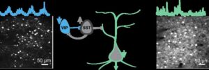
In particular, a class of inhibitory neurons known as vasoactive intestinal peptide (VIP) neurons, exhibited significantly reduced activity. VIP neuron activity is essential for normal learning, as these cells enable the brain to adapt and refine brain circuits during learning.
Reduced VIP neuron activity, the researchers reasoned, could be impairing the brain’s ability to function and learn properly. They sought to activate these cells to re-engage brain states that support learning. They tested this idea using optogenetics to stimulate VIP neurons.
“By activating the VIP inhibitory cell type, we gradually restored more normal activity patterns, and, very importantly, we also saw an improvement in the ability of the mouse to learn a motor task,” said Sonja Blumenstock, PhD, assistant project scientist at UC San Diego.
The results confirm VIP neurons as a key point of vulnerability in Huntington’s disease as well as a promising target for therapy. As to how this process works, the results suggest that modulating VIP neurons opens a “gate” that enables learning-related brain plasticity.
“This intervention restored more normal patterns of activity in the brain and improved movement in affected mice,” said Komiyama. “Importantly, the improvements persisted for days after stimulation ended, suggesting that the treatment triggered lasting beneficial changes in brain circuits rather than only temporary effects.”
The study provides important indications of where research could focus to normalize human brain function and facilitate brain recovery. Komiyama envisions a future scenario in which scientists could non-invasively activate the brain from outside the skull using novel approaches.
“Our study shows that despite the genetic defect, a precise intervention into the brain circuitry can lead to significant improvements in motor symptoms,” said Dudanova. “If we know which cells to target, we can retune the brain’s abnormal activity patterns. This gives hope for future therapies.”
The research also shows that corrections to specific brain circuit imbalances can restore function in a highly complex neurodegenerative condition, with similar potential in other disorders.
“We have come up with a way to allow the diseased brain to learn better,” said Komiyama. “The approach can improve behavior in diseased mice, and our hope is that a related approach will help people with impairment in their learning abilities.”
The post In Huntington’s Mouse, Optogenetic Activation of VIP Neurons Restores Brain Function appeared first on GEN – Genetic Engineering and Biotechnology News.
Biological Order Emerges from Tissue Boundaries, Drives Embryo Development
In a new study in Nature Materials titled, “Boundary geometry controls a topological defect transition that determines lumen nucleation in embryonic development,” researchers from European Molecular Biology Laboratory (EMBL) describe how interactions between tissue geometry impact development.
In an early-stage mouse embryo, cells of the epiblast are polarized and give rise to all major tissues. The team investigated the fundamental principles governing the behavior of polarized cells that are present in bulk and the impact of physical constraints at tissue borders. By focusing on how cellular orientations influence each other and their environment, the researchers built a minimal model that predicts how organization changes when interactions are altered.
“For me, as a physicist, I may know why something works, but it’s still kind of magic to see that it’s all true in messy biological systems,” said Pamela Guruciaga, PhD, postdoctoral researcher at EMBL and co-first author of the study. “It was also super interesting coming from a pure physics perspective to come up with a common language to work with biologists.”
In the cup-shaped epiblast, results showed different boundaries led to varying orientations for epiblast cells. When the boundary was lined with the extracellular matrix, the cells oriented perpendicularly. In contrast, when the epiblast was in direct contact with a neighboring tissue without a matrix, the cells aligned parallel to the boundary. The researchers found that the combination of these two orientations result in the appearance of structures, known as “topological defects.”
“These are points in space where it is undefined in which direction an object should point,” explained Guruciaga. “For example, if a set of arrows is arranged in a starburst pattern, the center is a point where all directions are equivalent. These points are super relevant because they are very robust; you cannot easily destroy them.”
To directly test whether the boundary shape controls the number of defects, the authors altered the geometry of the epiblast. Perturbing embryo shape induced the formation of additional lumina at the predicted positions.
“What I find most exciting is that these results identify a very general physical principle,” said Anna Erzberger, PhD, group leader at EMBL and co-corresponding author of the study. “We show that geometry alone can determine orientation patterns in three dimensions, independent of the microscopic details of the system. That means shape itself can act as a robust control parameter—not just in embryos, but across a wide range of biological and physical systems.”
The post Biological Order Emerges from Tissue Boundaries, Drives Embryo Development appeared first on GEN – Genetic Engineering and Biotechnology News.
CAR T Cells Simultaneously Target Glioblastoma and Immune Cells
Scientists have identified a new molecular target for CAR T-cell immunotherapy to attack both glioblastoma cells and tumor-supporting macrophages at once. A study published today in Nature shows promising preclinical results that could allow this approach to overcome the limitations of previous attempts to target glioblastoma with CAR T cells.
“Our approach targets both the tumor and the environment that allows it to thrive,” said Sheila K. Singh, MD, PhD, professor of neuro-oncology and neurosurgery at King’s College London and McMaster University. “Instead of treating glioblastoma as only a mass of cancer cells, we need to think of it as a connected tumor-immune ecosystem. By going beyond the cancer cells alone, we are also targeting immune cells that help shield the tumor from treatment.”
Glioblastoma is an aggressive and lethal form of brain cancer where current treatments, including surgery, radiation and chemotherapy, only provide temporary benefits and are rarely able to prevent recurrence. Past attempts to develop CAR T therapies for glioblastoma have failed to produce sustained responses due to a number of challenges such as heterogeneous antigen expression, antigen loss, and microenvironmental barriers that treatments solely focusing on targeting the tumor cells have not been able to surmount.
In particular, tumor-associated macrophages have been shown to be key contributors to glioblastoma progression. While macrophages normally play an important role in the immune response against infections, glioblastoma can recruit and reprogram these immune cells to promote tumor growth, suppress the immune system, and resist treatment.
“CAR T therapy has been effective in some blood cancers, but translating that success to brain tumors has been difficult,” said Shan Grewal, an MD/PhD candidate at McMaster and co-lead author of the study. “Most approaches have focused on killing cancer cells alone. Our work suggests we may also need to dismantle the immune support system that helps glioblastoma survive.”
Using patient tumor samples, Singh’s team conducted multi-omic profiling studies that led to the identification of a promising target present both in glioblastoma cells and tumor-associated macrophages, called glycoprotein non-metastatic melanoma protein B (GPNMB). By engineering CAR T cells to target GPNMB, the researchers were able to attack glioblastoma tumors on two fronts and show potent antitumor activity in several preclinical models including patient-derived xenografts.
While more work will be needed before this strategy can be evaluated in clinical trials, the study introduces a new framework to identify immunotherapy targets that could potentially be applied to a wide range of solid tumors beyond glioblastoma.
Supporting this concept, a team at the University of Calgary has simultaneously published results in Nature Cancer from a first-in-human study using a similar approach in relapsed alveolar soft-part sarcoma (ASPS) and translocation renal cell carcinoma, two types of cancer that stably express GPNMB. In these patients, a CAR T-cell therapy directed against GPNMB was found to be safe and induced stable disease for up to three months, providing early clinical evidence supporting the feasibility of this therapeutic approach.
The post CAR T Cells Simultaneously Target Glioblastoma and Immune Cells appeared first on Inside Precision Medicine.
A Recap of the Inaugural Youth Mental Health Hub at SXSW London
In early June, SXSW London returned for its second year, gathering thousands of creatives, enthusiasts, entrepreneurs, and investors into the city to celebrate film, music, tech, and culture. As part of this year’s festival, the Child Mind Institute, in partnership with Wellcome, proudly presented the inaugural Youth Mental Health Hub – a week of programming dedicated to advancing solutions to one of the defining challenges of our time: the global youth mental health crisis. Through six thought-provoking sessions, leaders in clinical care, science, technology, policy, and media came together to explore how to strengthen prevention, improve early identification, reduce stigma, and build systems that meet young people where they are.
Here’s a look back at the inspiring conversations that took place throughout the week.
Beyond the Average: Understanding Vulnerability in the Digital Childhood Era

As artificial intelligence rapidly transforms the experience of childhood, experts explored how AI can both support and challenge young people’s mental health. The panelists discussed when and under what conditions young people may be most vulnerable as well as what systems we need to support them.
Moderator
Gary Wilson, Director of Research, Huo Family Foundation
Speakers
Catherine Sebastian, PhD, Head of Evidence for Mental Health, Wellcome
John Pickavance, PhD, Principal Data Scientist, Born in Bradford
Georgia Turner, Postdoctoral Research Associate, University of Cambridge
Michael Milham, MD, PhD, Chief Science Officer, Child Mind Institute
AI Is Already Shaping Childhood. Who Is Shaping AI? Balancing Innovation, Evidence, and Safety in Youth Mental Health

Youth are experiencing the impacts of AI earlier and more intensely than any previous generation has. This session explored the role of public leadership in anticipating harm before it becomes systemic — establishing guardrails, fostering digital resilience, and ensuring that innovation advances hand in hand with youth mental health and well-being.
Moderator
Sarah Aguiar-Borges, PhD, University of Cambridge
Speakers
Julia Gillard, former Prime Minister of Australia; Chair, Wellcome
Kanishka Narayan, UK Minister for AI and Online Safety
Giovanni Salum, MD, PhD, SVP, Global Programs, Child Mind Institute.
Youth Mental Health After Conflict: Healing, Resilience, and Rebuilding Systems

Experts shared insights on the unique mental health challenges facing children affected by war, displacement, and humanitarian crises. This session explored how societies can implement youth-centered systems grounded in prevention and use early identification to position youth mental health as a cornerstone of long-term recovery and resilience.
Moderator
Krupa Padhy, BBC Radio 4
Speakers
Dr. Mark Jordans, professor, Centre for Global Mental Health, King’s College London; Director of Research & Development, War Child
Emma Ferguson, mental health policy and advocacy specialist, UNICEF
Mohamed Ali, Director, Iftin Global
Dyslexia: Changing the Story

In a timely discussion, experts explored how dyslexia is currently understood in society, challenging current language and misperceptions that can impact a child’s confidence and mental health. Through a blend of personal experience and clinical expertise, the conversation focused on the need for evidence-based support and strengths-based approaches to help children and their families thrive.
Moderator
Kate Griggs, Founder, Made By Dyslexia
Speakers
Maggie Aderin, PhD, space scientist & educator; dyslexia advocate
Harold S. Koplewicz, MD, President and Medical Director, Child Mind Institute
Connection Continuum: Preventing Suicide and Combating Loneliness

Suicide is one of the leading causes of death among young people globally. This session gathered community, clinical, and digital leaders to explore what a more connected system of support looks like in practice. The panelists also discussed the important of recognizing warning signs, expanding access to evidence-based care, and prioritizing early intervention to help prevent youth suicide.
Moderator
Krupa Padhy, BBC Radio 4
Speakers
Victoria Hornby, CEO, Mental Health Innovations
Dean Perryman, Empty Chairs
Michael Milham, MD, PhD, Chief Science Officer, Child Mind Institute
Does Mental Health Science Funding Need a New Paradigm in the Age of AI?

With technology evolving faster than the science designed to understand it, experts examined how research, philanthropy, and clinical leaders can work together to build the evidence, safeguards, and infrastructure needed to protect children’s mental health in the digital age.
Moderator
Chelsea Clinton, Vice Chair, Clinton Global Initiative
Speakers
Miranda Wolpert, Director of Mental Health, Wellcome
Margaret Laws, President & CEO, HopeLab
Daria Bukhman, Co-Founder and Chair, Bukhman Philanthropies
Harold S. Koplewicz, MD, President & Medical Director, Child Mind Institute
The post A Recap of the Inaugural Youth Mental Health Hub at SXSW London appeared first on Child Mind Institute.
Mammographic AI Adds Predictive Power to Breast Cancer Risk Models
A study by Kaiser Permanente researchers has shown that integrating mammographic AI with polygenic risk scores and clinical risk models can improve breast cancer risk stratification, guiding both personalized breast cancer screening and chemoprevention.
The study, published in the Journal of the National Cancer Institute, is one of the largest and most diverse to evaluate the ability of the three approaches to predict breast cancer risk.
“Our goal is to improve our ability to assess a woman’s breast cancer risk so we can personalize breast cancer screening recommendations,” said lead author Vignesh Arasu, MD, PhD, a radiologist and research scientist at the Kaiser Permanente Division of Research. “Our study shows that each of the approaches identifies a distinct group of women, and that when all three risk tests are used, we increase our ability to differentiate high-risk and low-risk women and provide more personalized screening recommendations.”
The study included 82,957 women (75% non-Hispanic White, 9% Asian, 7% Latina, and 4% Black) who enrolled in the Kaiser Permanente Research Bank between 2003 to 2020. All the women had a recent negative mammogram, and none had previously been diagnosed with breast cancer or had a genetic mutation known to increase breast cancer risk.
The researchers calculated each woman’s breast cancer risk using three different approaches: The Mirai mammography AI risk score, which looks for risk-related imaging biomarkers; the Breast Cancer Surveillance Consortium version 3 clinical risk score, which considers factors such as age, race or ethnicity, family history of breast cancer, breast density, and body mass index; and the 313-SNP polygenic risk score (PRS) that assesses risk based on the presence or absence of 313 breast cancer-associated single nucleotide polymorphisms.
During 10 years of follow-up, 2471 (3%) women were diagnosed with invasive breast cancer or ductal carcinoma in situ.
Arasu and team report that the C-index for breast cancer prediction when all three risk scores were combined was 0.70.
“This means that if you take any women who actually will get breast cancer in the future and pass her information to a risk model, that model will say she has a higher risk about 70% of the time relative to women who won’t get cancer,” Arasu explained.
The C-index for the combined model was significantly higher than that for individual models with only the clinical risk score (0.62), which is used by most clinical practices, the PRS (0.61), or the Mirai score (0.66).
The C-index for the triple model was also significantly higher than that for a model that combined the clinical and the polygenic risk scores (0.66).
The increase in the C-index of 0.04 “represents a moderate but meaningful improvement in discrimination when incorporating all three risk domains compared with the clinical plus polygenic model alone,” Arasu told Inside Precision Medicine.
He added: “The C-index reflects overall model performance across all possible risk thresholds and is commonly used as an initial, global assessment when integrating new predictors, such as AI-derived measures, to gauge their added value. However, future work will focus on defining clinically actionable risk thresholds to better characterize how these improvements translate into real-world tradeoffs between benefit and harm.”
Importantly, the improvements in risk prediction were consistent over time and across four common self-reported racial/ethnic subgroups of Asian, Black, Latina, and White women, which the authors say indicates that incorporating mammographic AI and ancestry-adjusted PRS into clinical risk prediction models may benefit all women.
The study also found that among the women at highest risk for developing breast cancer, the clinical risk score alone identified 19% of the women who went on to develop breast cancer over a 10-year period while the combined model identified 26% of these women.
Although all three model types are becoming increasingly accessible and part of care, Arasu cautioned that “more research is needed to see the benefit of using all three scores before a program that uses all three should be implemented.”
He added: “We already are planning the next research step, which will be to study the combined model in the on-going national WISDOM clinical trial in the next 1-2 years. The WISDOM trial already uses a PRS and clinical risk score to assess risk. Now, we will be adding mammographic AI.”
“By identifying more accurately which women are truly at high-risk, we hope to find more breast cancers as early as possible, when they are most easily treatable,” said Arasu. “For women who are very high risk, there is also the potential to discuss, in addition to annual mammography, risk reduction with a medication, such as tamoxifen.”
The post Mammographic AI Adds Predictive Power to Breast Cancer Risk Models appeared first on Inside Precision Medicine.
Stockholm3 Blood Test Detects More High-Risk Prostate Cancers Early
A new blood test may help address one of the key challenges in prostate cancer detection—identifying aggressive forms of the disease early on. According to research led by a team at Karolinska Institutet, the Stockholm3 blood test detected more clinically significant cancer cases than the well-established, but problematic, Prostate Specific Antigen (PSA) screening test.
The study appears in the Annals of Internal Medicine. Swedish researchers collaborated with teams from Europe and the U.S. on this work.
Prostate cancer is one of the top cancers among men globally, with an estimated 1.5 million new cases and 397,000 deaths annually. PSA testing has long been used for early detection. But, although PSA is prostate-specific, it is not cancer-specific. Elevated PSA levels can be caused by benign conditions as well as cancer and up to 50% of diagnosed aggressive prostate cancers are in men with low PSA values—below today’s cutoffs of PSA 3 ng/ml or PSA 4 ng/ml.
“There are a number of tests in development to improve on PSA alone. A lot of them, like Stockholm3, are fairly ingenuous and do a good job,” Mark Pomerantz told Inside Precision Medicine. He is a medical oncologist at the Dana-Farber Cancer Institute in Boston. Until one of these newer tests emerges as a winner, though, MRI is the gold standard for evaluating men with high PSA. “With MRI we can see the prostate in some detail,” Pomerantz said, “But it is expensive.”
The Karolinska-led researchers analyzed data from 12,670 men aged 50–74 from the population-based STHLM3-MRI study, which compared MRI-targeted and standard biopsy in men with elevated PSAs. In this more recent Annals study, the men were tested first with both PSA and Stockholm3 and followed for two years via national cancer registries, which allowed researchers to also identify cancer cases missed during the initial screening.
Stockholm3 detected 90 percent of aggressive cancer cases, compared to 74 percent for PSA.
“The test incorporates plasma protein biomarkers, genetic risk information from a polygenic risk score, and clinical factors such as age, family history, and prior biopsy history,” the study’s lead author Thorgerdur Palsdottir, told Inside Precision Medicine. Palsdottir is a researcher at the Department of Medical Epidemiology and Biostatistics, Karolinska Institutet.
He added, “Together, these components provide a more comprehensive estimate of a risk of harboring clinically significant prostate cancer than PSA alone.”
The components, he explained, map onto distinct biological axes rather than a single one. The kallikreins (PSA, free PSA, hK2) reflect prostate epithelial and tumor secretory activity, and the free-to-total PSA relationship helps separate benign enlargement from cancer. The polygenic score and family history capture inherited susceptibility—a man’s baseline predisposition—rather than anything about a tumor. GDF-15 is a stress-response marker that has been associated with more aggressive disease across several cancers.
During the study follow-up, 443 men were diagnosed with clinically significant, i.e. aggressive, prostate cancer. Stockholm3 missed significantly fewer serious cancer cases than PSA, while the proportion of men incorrectly classified as high-risk was similar between the tests.
“These results point toward a potential change in how prostate cancer screening can be conducted. A more precise blood test could enable earlier detection of aggressive disease while reducing the number of unnecessary follow-up examinations and procedures,” said Palsdottir.
He adds that longer-term follow-up is needed to fully assess the effects on mortality and long-term outcomes. “The next important step is to evaluate longer-term outcomes, including disease progression, metastatic disease, and prostate cancer mortality.”
The post Stockholm3 Blood Test Detects More High-Risk Prostate Cancers Early appeared first on Inside Precision Medicine.