
Background: Digital health offers opportunities to facilitate symptom assessments and communication for children with cancer, particularly after discharge. However, access to these tools must be established to ensure that they effectively support the user. PicPecc (Pictorial Support in Person-Centered Care for Children) is a mobile health tool developed to enable children to remotely assess symptoms and communicate with health care professionals. Understanding access to PicPecc is essential for evaluating its use in pediatric oncology. Objective: The aim was to test a digital intervention with PicPecc in pediatric oncology care through the lens of access to technology. Methods: This study uses a triangulation approach to determine access to digital technology through an intervention, PicPecc outside hospital. Fourteen children (6‐17 y), 5 parents, and 6 nurses from 2 pediatric oncology units in Sweden participated. Children were encouraged to use PicPecc for 2 weeks (achieving a median of 14, IQR 9.75-16 days) following hospital discharge to assess pain, nausea, sleep disturbances, and feelings using an assessment scale, pictures, personal notes, and a chat function. Nurses monitored assessments and responded via the administrative interface. Access was analyzed through interviews and an instrument, and by recording the consumption of PicPecc. Data analysis was based on the 5 dimensions of access (availability, accessibility, accommodation, affordability, and acceptability). Results: The intervention, PicPecc outside hospital, supported availability by enabling children to communicate symptoms in a safe and structured way. Children and parents mentioned feeling safe when they were discharged from the hospital, and nurses perceived it as a valuable complement to follow-up after discharge. PicPecc outside hospital was generally accessible, although initial challenges with log-in procedures related to the PIN code were common. Barriers related to accommodation included interpreting the scale and obtaining an overview of assessments. Affordability was high, as internet access and device availability were not barriers; however, children’s motivation varied depending on symptom burden. Acceptability was strong among children up to 12 years of age, who appreciated the design and gaming function, while the older children found the visual design less age-appropriate. Conclusions: Access to the mobile health tool, PicPecc outside hospital, appears promising for supporting remote symptom assessment in pediatric oncology, particularly among children up to 12 years of age. However, identified barriers, such as motivational factors and integration into the health care system, need to be addressed.
<img src="https://jmir-production.s3.us-east-2.amazonaws.com/thumbs/0e5b1596261b54081796260d31e302f1" />
Experiences With Technology Among Adults Aging With HIV Engaged in an Online Community–Based Exercise Intervention Study: Longitudinal Qualitative Descriptive Study and Secondary Data Analysis
Background: As individuals with HIV live longer, many now face the health consequences of aging and multimorbidity, known as disability. Exercise can mitigate disability; however, engagement in exercise among adults living with HIV varies. Technology-based interventions, such as telerehabilitation, may help mitigate geographical, financial, and time barriers to community-based exercise (CBE). However, little is known about the experiences with technology uptake and usage among adults living with HIV. Understanding these experiences is essential to inform the design of inclusive, accessible, and sustainable online interventions. Objective: This study aimed to describe experiences with technology uptake and usage among adults aging with HIV participating in a 6-month online CBE intervention and explore how these experiences changed over time, from baseline to postintervention. Methods: We conducted a longitudinal qualitative descriptive study and secondary analysis using interview data from adults living with HIV who were engaged in a CBE intervention study in Toronto, Canada. Participants engaged in a 6-month online CBE intervention consisting of thrice-weekly exercise supervised biweekly through online personal coaching sessions, weekly group exercise classes, and monthly self-management education sessions (via Zoom). The technology used included Zoom software and a webcam, as well as the Sweat for Good YMCA app and the YMCA Virtuagym website; participants wore a wireless physical activity monitor (Fitbit Inspire 2) throughout. Participants completed interviews at baseline and postintervention. We conducted a group-based content analysis of interview transcripts, focusing on digital access, setup, usage, and perceptions of technology. Questionnaire data describing digital literacy and access to technology provided additional context to the interview data. Results: Eleven participants completed at least one interview. We analyzed 19 interview transcripts from 11 participants (women: n=6, 55%; men: n=5, 45%; median age 52, IQR 45-60 y). Experiences with technology uptake and usage among adults aging with HIV were characterized by four components: (1) preparations for technology (technology setup), (2) interactions with technology (preferences for different types of technology, preferences for mode of delivery, and ease of usage), (3) facilitators and satisfaction with technology (facilitators to technology uptake and usage and satisfaction with technology), and (4) challenges and frustrations with technology (barriers to technology uptake and usage and frustrations with technology). Experiences with technology across participants were influenced by intrinsic contextual factors (prior exposure to technology) and extrinsic contextual factors (COVID-19 pandemic and technological and social support). Conclusions: Experiences with technology among adults aging with HIV engaging in an online CBE intervention varied from increasing ease of use to increasingly burdensome over time. Results highlight the need to incorporate personal preferences and ongoing technological support when implementing online CBE with adults aging with HIV.
<img src="https://jmir-production.s3.us-east-2.amazonaws.com/thumbs/0a8d6a5175ae1cbe3aafc0c9c2827f3c" />
STAT+: Medicare takes another swing at 340B cuts to hospitals
Medicare wants to slash payments to hospitals for drugs acquired through the 340B drug discount program by more than a third beginning next year, after the agency said its surveys found some patients paid more for the drugs than the hospitals did.
Under a proposal released Thursday, Medicare would pay hospitals for 340B drugs at their average sales price minus 33.4%, dramatically less than they’re getting currently, which is that price plus 6%. The provision, part of a proposed rule on hospital outpatient payments, represents the latest swing at what’s become a hotly debated drug discount program, viewed by some as a lifeline for safety-net hospitals and by others as a profit center for wealthy health systems.
The proposal drew swift condemnation from groups representing nonprofit and academic hospitals, who said it would disproportionately harm safety-net providers. That’s because only these nonprofit facilities are eligible for 340B, while for-profit hospitals are not. Medicare’s proposed rule shows a 7.4% pay increase to for-profit hospitals under the 340B adjustment.
Synthetic Organizers Aid Creation of Reproducible Kidney Organoids from Stem Cells
University of Southern California (USC) researchers have paired a biological discovery with an engineering feat to create more faithful, reproducible kidney organoid structures, grown from human pluripotent stem cells (hPSCs). By mapping the developing human kidney, the scientists identified a previously unrecognized developmental axis that helps organize the kidney’s nephrons, which are their filtering units. The team then engineered Wnt-secreting “synthetic organizer” cells to recreate aspects of this developmental environment in organoids.
Their advance makes the organoids more reliable models for studying disease and evaluating potential therapies, while supporting long-term efforts to generate transplantable kidney tissue. “It is important that we’re starting to get good reproducibility from organoid models that can lead to robust preclinical models of cell function and disease to benefit patients,” said Nils Lindström, PhD, assistant professor of stem cell biology and regenerative medicine at the Keck School of Medicine of USC. Lindström is co-corresponding author of the team’s published report in Science, titled “Patterning human kidney organoids with synthetic Wnt-secreting organizers.” In their paper, the researchers reported, “Our findings link a spatial organizing geometry in the developing human kidney to controllable engineering in vitro.”
“Stem cell–derived organoids have emerged as systems for modeling organ development and generating complex tissue structures in vitro,” the authors wrote. Over the past decade, organoid work has relied on cells’ ability to self-organize into tissue-like structures, often in response to adding chemicals and proteins that act broadly in the whole organoid. “Although this capacity enables organoids to recapitulate many developmental programs, it limits experimental control over tissue architecture, often producing structures that vary between cultures and are difficult to engineer reproducibly,” the team continued. “Understanding how to impose spatial patterning in organoid systems is therefore an important challenge.”
In embryos, spatial patterning is often organized by localized signalling centers, known as developmental organizers. But how organizing geometry is controlled in the developing kidney, and whether it can be recreated in vitro, hasn’t been known.
For their reported study, the team combined spatial transcriptomics of the developing human kidney with synthetic engineering. “We mapped this organizing geometry in developing human kidneys and tested whether a minimal cue of localized WNT signaling could restore spatial control of nephron patterning in organoids,” they explained.
The project began by making tools to copy developmental signals. Postdoctoral researcher Fokion Glykofrydis, PhD, in the Morsut lab, engineered a “synthetic organizer” cell that secreted a Wnt protein that their spatial transcriptomics and other analyses indicated was involved in spatial patterning during kidney development. Graduate student Connor Fausto from the Lindström lab proposed an experiment to test how this Wnt-secreting cell would affect organoid nephrons.
The experiments revealed that the synthetic organizer enabled two key processes essential for building organs: controlling the identity of cells and influencing the shape of developing structures. The synthetic organizer serves as a localized and targeted source that secretes controllable amounts of specific Wnt proteins within the organoid itself. These are key signals that help shape the developing kidney. This creates a signaling environment much more similar to a naturally developing kidney and gives researchers a way to control where and how kidney structures form.
Co-corresponding author Leonardo Morsut, PhD, associate professor of stem cell biology and regenerative medicine, and biomedical engineering at the Keck School of Medicine and USC Viterbi School of Engineering, said, “With our approach, we are trying to control self-organization, and work with it as opposed to try to completely override it.”
Lindström expected Wnt to trigger nephrons to change their identity into cells capable of forming connections with the urine drainage system. What surprised him was that the nephrons also changed shape and elongated toward the source of the Wnt signal, which doesn’t happen when signals are delivered uniformly to the whole organoid. Compared with the developmental process seen in traditional kidney organoids, this elongation toward the Wnt source is more similar to what happens in a naturally developing kidney.
“A single, localized signal did two things at once. It changed what the cells became and physically pulled the tubules toward the source,” Lindström said. “You would not see that with a uniform chemical bath of signals.” Engineered WNT-secreting cellular organizers introduced into kidney organoids restored organizing geometry, the authors noted, “… biasing distal nephron differentiation and orienting nephron morphogenesis toward the signal source, which demonstrates that developmental signaling geometry can be reconstructed synthetically to control tissue patterning.”
The team identified a previously unrecognized axis, a direction along which the developing kidney organizes itself. Developmental biologists have long known about the nephron’s classic “proximal-distal (PD) axis,” which runs from its blood-filtering end to its urine-drainage end. The new axis is defined instead by how close each part of the nephron sits to the collecting duct, the tube system that drains urine and releases Wnt signals during development. Those signals tell the nephron what shape to take and which way to point.
“The study shows that there’s an undiscovered axis that sets up how a nephron looks and forms,” said Lindström. “It’s not every day that you find something new in human development at that level.”
Most kidney organoids contain only nephrons and lack the collecting duct that supplies this local Wnt signal, so they have no such axis and organize in a radially symmetrical pattern. By mapping how kidney cells respond to Wnt at specific locations in the developing kidney, the team recreated that environment in organoids with the synthetic organizer, producing structures that are both more developmentally faithful and more reproducible.
“Introducing tunable WNT-secreting synthetic organizers (SOs) in organoids restored canonical WNT responses, biased distal nephron differentiation, and oriented nephron morphogenesis toward the WNT source,” the investigators stated in summary. The combined results, they suggested, “… demonstrate that the spatial geometry observed in vivo can be reconstructed synthetically to control early nephron patterning and morphogenesis … Synthetic organizers provide a modular way to restore missing spatial interactions without reconstructing the entire collecting duct lineage, complementing approaches that rebuild collecting duct–to–nephron cellular interactions.”
For Morsut, the synthetic organizer is one of several tools his lab is building to control how tissues form, and the one he is most excited about, because it steers development in a way that is powerful but not intrusive. “The synthetic organizer is just a little cluster of cells that don’t build anything themselves,” said Morsut. “But they produce a powerful field that aligns the stem cells and gives them a direction.”
Synthetic organizers offer a modular strategy to reintroduce spatial signaling interactions that are often absent in conventional organoid cultures, the team suggested. “This approach should be broadly applicable to other organoid systems in which spatial signaling environments play instructive roles during development, providing a framework for linking developmental biology with the rational engineering of tissue architecture.
Aligning cells is something embryos do repeatedly as they build themselves, Morsut noted, and the study shows it can now be put to work in an engineering setting, steering the process toward a desired outcome. “At the beginning of my talks, I always show a video of embryonic development,” said Morsut. “You start from a single cell, and you get to a complete organism, and that’s as close to magic as it gets. Now, we open a possibility of controlling this magic technology for building organs. This study shows that we can do that, and I’m excited to see what others will do in other contexts.”
The post Synthetic Organizers Aid Creation of Reproducible Kidney Organoids from Stem Cells appeared first on GEN – Genetic Engineering and Biotechnology News.
ASMS 2026: Solving Proteomics’ Next Bottleneck
At the 74th American Society for Mass Spectrometry (ASMS) Conference in San Diego, the obvious story was hardware. Vendors showcased faster acquisition, higher sensitivity, alternative fragmentation, spatial workflows, and software ecosystems. New or highlighted platforms and workflows came from Waters, Thermo Fisher Scientific, Sciex, Bruker, Biognosys, and Evosep.
But after several days of talks, posters, hallway conversations, and interviews with senior figures in mass spectrometry (MS)-based proteomics, the deeper story was not simply that instruments are getting better. The field is beginning to look past the instrument. The mass spectrometer is still central, but the question is shifting: what has to happen around it for proteomics to become clinically useful, scalable, trusted, and routine?
Beyond the instrument
Jennifer Van Eyk, PhD, professor of cardiology, biomedical sciences, pathology, and laboratory medicine, and director of the Advanced Clinical Biosystems Research Institute at Cedars-Sinai Health Science University, put it most directly: “I think mass spec is no longer the limitation. We have the sensitivity, the throughput, and the accuracy at discovery and targeted levels.”

That is a remarkable statement in a field long defined by instrument performance. Van Eyk was not saying that MS innovation is finished. She pointed to continuing gains in quantitation, protein structure, conformational analysis, post-translational modifications (PTMs), top-down proteomics, and protein dynamics. But for clinical impact, she argued, the next bottlenecks are increasingly sample preparation, data analysis, standardization, harmonization, and quality control.
Joshua Coon, PhD, professor of biomolecular chemistry at the University of Wisconsin-Madison and the Pyle Chair at the Morgridge Institute for Research, saw instrument speed as the force opening new applications. Faster scanning mass analyzers are allowing deeper proteome coverage, more post-translational modification (PTM) measurements, and shorter runs. Ryan Kelly, PhD, professor of chemistry and biochemistry at Brigham Young University, framed the same shift as a throughput problem. “Now the mass spec is so fast that we need to figure out how to feed it faster,” he said. In plasma proteomics, Coon said, faster instruments, nanoparticle-based enrichment, and improved chromatography are moving the field from hundreds

toward thousands of detectable proteins in blood.
John R. Yates III, PhD, the John Lytton Young Endowed Chair in the department of integrative structural and computational biology at Scripps Research, highlighted electron activation dissociation methods and the possibility that high-throughput workflows could push MS deeper into plasma and population-level studies. He described targeted affinity platforms as powerful for “known knowns” because they measure targets defined in advance. “But with mass spectrometry,” he added, “you can look for unknown unknowns, which is where the gold lies.”

The point cuts to the heart of where the field now stands, and a recurring ASMS tension. The future of proteomics is not a choice between platforms. It is a division of labor. Targeted affinity technologies have become central to large-scale plasma proteomics and population studies. MS remains uniquely powerful for unbiased discovery, tissue proteomics, complex sample matrices, protein modifications, structural diversity, and biology that is not yet named.
From depth to trust
If the first era of modern proteomics was about seeing more, the next may be about measuring better. Devin Schweppe, PhD, assistant professor in the Department of Genome Sciences at the University of Washington, described the current moment as a “duality.” Instruments can now deliver deep coverage, and computational tools are making interpretation faster. Together, he said, they are creating “a comfort level with trusting the data.”

Trust came up repeatedly. For discovery biology, a strong signal can be enough to generate a hypothesis. For clinical practice, it is not. Van Eyk said clinical-grade assays are “way harder than people think they are.” A research study can iterate. A clinical assay has to deliver the same measurement today, in five weeks, in six months, and years later. Once a test is locked, “you can’t go, ‘Oh no, we should have had this extra protein in there,’” she said. “It’s done.”
This distinction matters across assay types. Targeted MS methods such as multiple reaction monitoring (MRM) and parallel reaction monitoring (PRM) can provide absolute quantification, but only for preselected proteins. Data-independent acquisition (DIA), meanwhile, has moved discovery proteomics closer to translation by improving reproducibility and scalability. DIA is still often used for relative quantification, but its ability to capture patterns across tens or hundreds of proteins may become important as clinical decision-making moves beyond single biomarkers and reference intervals.
The field is responding to these demands. David Kotol, PhD, R&D manager at ProteomEdge, discussed an independently validated nine-protein plasma panel designed to improve emergency department triage and imaging decisions for patients with suspected venous thromboembolism, compared with D-dimer alone.

Kotol described a shift “from relative protein measurements toward robust, multiplexed absolute quantification.” He emphasized stable isotope-labeled protein standards added early in sample preparation to monitor digestion efficiency, downstream analytical variation, and multi-peptide quantification. These standards cannot remove variation introduced during sample collection, handling, or storage. But they can make the analytical workflow more transparent and transferable.
The clinical gap
Mathieu Lavallée-Adam, PhD, associate professor in the department of biochemistry, microbiology and immunology and director of the specialization in bioinformatics at the University of Ottawa, gave the least glamorous answer to what still blocks clinical translation. “My answer is going to be boring,” he said. “It’s going to be education.”

Lavallée-Adam argued that many clinicians and biomedical researchers still do not fully understand what modern MS can do. Too often, the outside view is still: give me a list of differentially expressed proteins. But MS-based proteomics has moved beyond lists, into proteoforms, structural information, PTMs, protein dynamics, and flexible acquisition. “We’re past that now,” he said. “The main barrier is our inability to communicate the possibilities that we offer.”
Sasha Singh, PhD, assistant professor of medicine at Harvard Medical School, associate scientist at Brigham and Women’s Hospital, and director of proteomics research at the Center for Interdisciplinary Cardiovascular Sciences (CICS), described this translation role from inside a hospital environment. “That’s actually my role at the hospital,” Singh said. “I am a liaison between the technology and the application scientist.”

The translation is becoming harder because proteomics is diversifying. End users often need to distinguish among discovery MS, which can provide broad relative quantification; targeted MS, which can provide absolute concentrations for selected proteins; and targeted affinity proteomics, which can scale well for plasma cohorts but is limited by predefined assays and available binding reagents. Singh added that different technologies may produce profiles that do not fully overlap. Rather than treating that as a failure, she suggested it reveals something real: the circulation contains many subproteomes, and different technologies enrich different views.
AI with guardrails
No 2026 conference escapes artificial intelligence (AI), and ASMS was no exception. But the mood among the researchers was cautious rather than breathless.
Lavallée-Adam said agent-based AI was dominating conversations in his part of the field. The dream is seductive: put a sample on an instrument, ask an AI agent to maximize protein identifications or optimize a method, and let it select the best protocol. But he drew a clear line between potential and reality. “Are such agents really driving change? It’s unclear at this point,” he said. “I think it’s unproven.”
Still, AI-assisted acquisition strategies are entering workflows. Lavallée-Adam’s group works on real-time MS data acquisition, where software analyzes data as it is acquired and adapts the run to the biological question. Instead of measuring the same abundant proteins repeatedly, the system can decide it has seen enough and move on to new targets. In that sense, AI becomes less a magical oracle than an instrument assistant.
Faster instruments are generating more data, and faster analysis is needed to keep up. Schweppe also argued that open-source tools remain essential because they let laboratories build on one another’s work rather than rebuild it.
More than abundance
Much of the clinical proteomics effort is focused on plasma because it is minimally invasive and suitable for screening, longitudinal sampling, and routine monitoring. But even in blood, researchers are learning that plasma is only part of the story.
Roman Fischer, PhD, associate professor and head of the Discovery Proteomics Facility at the Target Discovery Institute, University of Oxford, pushed the conversation back toward biology. Plasma alone does not capture the full circulating system, he noted. Peripheral blood mononuclear cells, extracellular vesicles, microvesicles, and other compartments may contain disease-relevant information that conventional workflows miss. “We have to be more sophisticated in addressing the compartments of the blood,” Fischer said.

He also pointed to the proteoform problem. A single gene can give rise to many transcripts, isoforms, modified proteins, and glycosylated forms. These differences may affect activity, localization, disease pathways, and therapy response. Capturing that diversity is not possible with targeted affinity assays alone. It requires deeper characterization of the proteome, not only quantification.
Yates offered a clinical example. His group has been developing protein-footprinting approaches that can detect conformational changes in proteins in blood. In one transthyretin amyloid cardiomyopathy project, he said, abundance alone was not the answer. The important signal was how the protein folded or misfolded. That kind of assay moves proteomics beyond proteins going up or down, into structural disease biology.
Van Eyk’s work on remote sampling devices pointed to another future: patient-collected blood samples that make longitudinal cardiovascular studies easier, more inclusive, and better matched to real clinical questions.
In the background was a broader translational arc: discovery, verification, clinical validation, health economics, and access. Plasma proteomics highlights included Lekha Sleno, PhD, professor at Université du Québec à Montréal, who is combining nanoparticle enrichment with isotope-enabled targeted proteomics, and a CinderBio breakfast seminar featuring Fredrik Edfors, PhD, assistant professor at KTH Royal Institute of Technology and SciLifeLab, and Simion Kreimer, PhD, senior research project advisor in the Proteomics and Metabolomics Core at Cedars-Sinai Health Science University.
The seminar focused on accelerated plasma proteomics, rapid digestion workflows, stable isotope standards, Human Protein Atlas resources, and faster enzyme workflows that can reduce lead times. The common message was that sample preparation, quantification, and validation may become as decisive as instrument resolution.
The next bottleneck
ASMS 2026 was not short on technical spectacle. High-resolution instruments, electron-based fragmentation, narrow-window DIA, rapid acquisition, MS imaging, top-down workflows, and AI-enabled software all had their moment. But the most interesting conversations were less about spectacle than maturity.
Proteomics is no longer trying only to prove that it can see more. It is trying to prove that it can measure consistently, explain biology more deeply, support drug development, fit into clinical laboratories, and eventually improve patient decisions.
That means the next bottleneck is distributed across the ecosystem: sample preparation, standards, software, education, reimbursement, clinical menus, regulatory validation, open tools, and the ability to translate technical power into something a clinician can use.
Longer term, integrated proteomics, other omics, imaging, clinical data, and AI may support not only single biomarkers, but interpretable molecular patterns, longitudinal trajectories, and digital-twin-like models of patient biology.
The field spent decades making proteins visible. The next challenge is making proteomic measurements dependable enough to act on.
The post ASMS 2026: Solving Proteomics’ Next Bottleneck appeared first on GEN – Genetic Engineering and Biotechnology News.
Automated Optic Disc Tilt Classification in Fundus Photographs Using Segmentation and the Elliptical Ratio: External Clinical Validation Study
Background: Optic disc tilt is a morphological change in myopic eyes that complicates clinical interpretation and artificial intelligence (AI)–based analysis of fundus images. Accurate detection of optic disc tilt is necessary to avoid misinterpretation of disc morphology and enhance diagnostic reliability across different disease types. Objective: This study developed and externally validated an end-to-end AI-based pipeline for optic disc segmentation and quantitative tilt classification in color fundus photographs (CFPs), offering an objective alternative to manual segmentation and subjective clinical assessments. Methods: We trained a nnU-Net–based optic disc segmentation model on the Standardized Multi-Channel Dataset for Glaucoma (SMDG; n=3103 CFPs) and externally validated it on the Samsung Medical Center (SMC) dataset (n=2448 CFPs from n=1370 patients). Model generalizability was assessed using both a fixed 80:20 random split and 5-fold cross-validation. Tilt was classified using the ratio of the long-axis diameter to the short-axis diameter, with a ratio of ≥1.3 indicating tilt. Segmentation performance was evaluated using the Dice similarity coefficient, intersection over union, and pixel accuracy on the SMDG dataset and using the clinical acceptance rate determined by 2 independent ophthalmologists on the external SMC dataset. Results: Using the SMDG dataset, nnU-Net achieved consistently high performance, with mean Dice similarity coefficients of 0.956 (SD 0.042) across 5-fold cross-validation and 0.961 (SD 0.055) for the best-performing single-fold model across 8 datasets. On the SMC dataset, 2 independent expert reviews yielded mean clinical acceptance rates of 98.61% and 98.86% across disease types, with acceptance rates ranging from 81.63% and 93.88% for edema to 99.59% and 99.17% for pallor, respectively. Tilt was detected in 7.5% (186/2448) of images, with rates of 9.7% (118/1215) for normal images, 3.9% (35/894) for glaucoma, 7.8% (19/241) for pallor, and 14.2% (14/98) for edema. Segmentation errors were observed in 1.39% (34/2448) and 1.14% (28/2448) of cases by the 2 reviewers, mainly due to edema-related swelling, peripapillary atrophy, and vessel confusion. Conclusions: Our pipeline provides objective and reproducible detection of optic disc tilt in CFPs, with strong generalizability to clinical images. By replacing manual segmentation and subjective assessments, the pipeline supports tilt-aware AI diagnostics and scalable screening for myopia-related conditions, with future refinements needed to address edema-related challenges.
<img src="https://jmir-production.s3.us-east-2.amazonaws.com/thumbs/9d238734c91be565746472427448cd3f" />
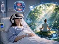
Effects of Virtual Reality on Postoperative Pain Management Following Minimally Invasive Gynecologic Surgery: Randomized Controlled Trial
<strong>Background:</strong> Postoperative pain and anxiety remain common concerns after minimally invasive gynecologic surgery despite advances in surgical techniques and analgesic strategies. Virtual reality (VR) has been investigated as a potential nonpharmacological intervention for pain management; however, evidence in gynecologic postoperative settings is limited. <strong>Objective:</strong> This study aims to evaluate the efficacy and safety of VR technology compared with standard postoperative analgesia for pain and anxiety management in patients undergoing minimally invasive gynecologic surgery. <strong>Methods:</strong> This randomized controlled trial was conducted at Sun Yat-sen Memorial Hospital of Sun Yat-sen University in China. A total of 131 patients undergoing laparoscopy or combined hysteroscopy for benign gynecologic diseases were randomly assigned in a 1:1 ratio to either a VR group (n=68) or a control group (n=63). All patients received a standardized general anesthesia protocol intraoperatively. The control group received conventional analgesic therapy after surgery, and the VR group received a 20-minute VR intervention 6 hours postoperatively. The pain and anxiety levels were evaluated using a visual analog scale at 6 and 7 hours postoperatively. The primary outcome was the change in pain scores between 6 and 7 hours. Secondary outcomes included maximum pain score, anxiety score changes, length of hospital stay, hospitalization costs, and occurrence of adverse events. Analyses were performed according to the intention-to-treat principle. <strong>Results:</strong> There was no statistically significant difference in the primary outcome between the VR and control groups (mean difference 0.169, 95% CI −0.271 to 0.608; <i>P</i>=.45). Similarly, no significant differences were observed in the maximum pain score (mean difference 0.839, 95% CI −0.101 to 1.779; <i>P</i>=.08), and no improvement was observed in the anxiety score (mean difference 0.042, 95% CI −0.365 to 0.449; <i>P</i>=.84). No significant differences were found in length of hospital stay, hospitalization costs, or incidence of adverse events, including dizziness, nausea, and vomiting (all <i>P</i>>.05). <strong>Conclusions:</strong> A single 20-minute VR intervention did not provide additional analgesic or anxiolytic benefit compared with standard postoperative care after minimally invasive gynecologic surgery. VR was well tolerated, and its role in postoperative recovery requires further investigation. <strong>Trial Registration:</strong> Chinese Clinical Trial Registry ChiCTR2400091244; https://tinyurl.com/4b92a9td
From Alliance to Nexus: Rethinking Digital Therapeutic Relationships
In traditional human psychotherapy, the therapeutic alliance (TA) is regarded as a fundamental factor that describes the client-therapist relationship, mainly due to strong evidence demonstrating its impact on treatment outcomes regardless of theoretical orientation. More recently, advances in artificial intelligence (AI) and other technologies have led to the emergence of the concept of digital TA, used to characterize the relationship between clients and AI-based therapeutic systems. This approach replicates human dynamics but overlooks key differences between human therapists and digital agents. Prematurely translating the concept of TA into the digital context fails to address issues such as the sycophantic tendencies of current systems and the inherent limitations of algorithmic interaction. We propose the digital therapeutic nexus, a framework that recognizes these differences and provides a set of structured criteria for categorizing digital interactions into 3 progressive levels. This Viewpoint argues that only at the highest level can parallels be drawn to the human TA and stratifies the main risks associated with each nexus level. Transitioning from the concept of alliance to that of a nexus offers a more precise conceptual basis for describing and evaluating digital therapeutic relationships, with implications for research, design, and the ethical development of AI-based mental health interventions.
<img src="https://jmir-production.s3.us-east-2.amazonaws.com/thumbs/eedf1536589b3edd0896d4525b7a411e" />
Blood circRNAs Can Predict Alzheimer’s Years Prior to Symptoms Onset
A set of blood-based circular RNAs (circRNAs) could change how we diagnose and monitor Alzheimer’s disease, providing a simple, noninvasive test that can detect the disease with remarkable accuracy and predict its progression years before symptoms appear.
Washington University School of Medicine researchers analyzed blood samples from 1,221 individuals, including people with Alzheimer’s disease and cognitively healthy participants, making it one of the largest investigations of blood circRNAs in Alzheimer’s to date. The findings, published in a Nature Medicine study, identified 34 circRNAs whose combined expression patterns accurately distinguished Alzheimer’s disease from healthy aging.
Unlike conventional RNA molecules, circRNAs form single-stranded closed loops that resist degradation and are abundant in the brain. Their stability and ability to cross the blood-brain barrier make them attractive candidates for blood-based biomarkers that reflect changes occurring in the brain.
The research indicated that a predictive model built from the 34 circRNAs achieved an area under the curve (AUC) of 0.945 for identifying biomarker-confirmed Alzheimer’s disease, outperforming the widely used plasma biomarker pTau217 (AUC 0.877). When circRNA measurements were combined with pTau217, diagnostic performance increased further to an AUC of 0.977.
Beyond diagnosis, the circRNA signature demonstrated exceptional ability to predict disease progression. Individuals with elevated circRNA scores were nearly three times more likely to progress to symptomatic Alzheimer’s disease than those with lower scores. The model also outperformed pTau217 in forecasting progression and remained highly specific for Alzheimer’s, showing limited predictive ability for other neurodegenerative disorders such as Parkinson’s disease, frontotemporal dementia, and dementia with Lewy bodies.
Importantly, the findings were independently replicated in two additional cohorts, including 551 participants from the Knight Alzheimer’s Disease Research Center and 1,767 participants enrolled in the Anti-Amyloid Treatment in Asymptomatic Alzheimer’s Disease (A4) study. This independent validation demonstrates that the circRNA signature is robust across multiple populations and study designs.
The researchers also found evidence that circRNA changes begin approximately two to four years before the onset of clinical symptoms, suggesting that these molecules may capture biological processes closely linked to the transition from silent pathology to cognitive decline. Such biomarkers could become increasingly valuable as disease-modifying therapies enter clinical practice, where monitoring ongoing neurodegeneration is just as important as detecting amyloid pathology.
The work builds on intellectual property protected by patent PCT/US2026/017857, “Blood Circular RNA as a Noninvasive Biomarker of Alzheimer’s Disease,” which Circular Genomics has licensed. The company, based in San Diego, California, is developing next-generation molecular blood biomarker diagnostics for precision neurology. The patent covers the use of blood circRNA signatures for the diagnosis and monitoring of Alzheimer’s disease and supports continued development of clinically deployable blood tests.
While the authors emphasize that larger prospective studies are still needed before widespread clinical implementation, the findings position blood circRNAs as a promising new class of biomarkers for Alzheimer’s disease. By combining high diagnostic accuracy with strong prediction of disease progression using a simple blood sample, circRNA-based testing could help identify patients earlier, improve clinical trial enrollment, and provide physicians with new tools to monitor disease over time.
The post Blood circRNAs Can Predict Alzheimer’s Years Prior to Symptoms Onset appeared first on Inside Precision Medicine.
Achieving operational excellence with AI
Frameworks like Lean Six Sigma and business process management (BPM) first gained traction because they promised clarity in the chaos—a structured way to bring order to messy, sprawling operations. Lean Six Sigma emphasized statistical rigor and quality control; BPM created end-to-end maps of how work should flow across departments. Both offered a repeatable way to embed habits of measurement, analysis, and accountability into day-to-day company culture.

But today, those time-tested playbooks are evolving as companies seek to embed AI into established process excellence methodologies. By some estimates, the market for AI-powered process optimization is projected to exceed $113 billion within the next decade. In one study, a full 88% of business leaders anticipated increasing investments into AI-infused process intelligence in the next 12 to 18 months.

Yet without the right foundations, many of those investments may not fully deliver on their potential. Companies that already operate with discipline have an edge. They can channel new tools into proven systems rather than bolting them onto shaky foundations. Organizations with mature process disciplines are also better positioned to translate AI ambition into real outcomes, as they are already accustomed to data-driven decision-making and process discipline—precisely the cultural foundation AI systems need to deliver value.
Simply put: AI can accelerate process excellence, but existing process excellence is what makes AI truly impactful. Technology and process are no longer separate levers, and only organizations that pull them together stand to realize the full value of both.
This content was produced by Insights, the custom content arm of MIT Technology Review. It was not written by MIT Technology Review’s editorial staff. It was researched, designed, and written by human writers, editors, analysts, and illustrators. This includes the writing of surveys and collection of data for surveys. AI tools that may have been used were limited to secondary production processes that passed thorough human review.