Nature Biotechnology, Published online: 16 June 2026; doi:10.1038/s41587-026-03195-0
Light-powered metabolism in the mammalian eye

Radically different
Nature Biotechnology, Published online: 16 June 2026; doi:10.1038/s41587-026-03195-0
Light-powered metabolism in the mammalian eye
Background: Although patient outcomes are improved by stroke rehabilitation, the suggested amount of therapy is rarely maintained. The COVID-19 pandemic aggravated this situation further due to disruptions in health care. One solution was the rapid and extensive transition to virtual care. A hybrid outpatient stroke telerehabilitation program (HOSTP) was introduced by St John’s Rehab—a tertiary rehabilitation hospital in Toronto, Ontario. The HOSTP integrated in-person and virtual care in an effort to alleviate long-standing obstacles that challenge stroke rehabilitation. Objective: This study explored health care providers’ (HCPs) experiences with the HOSTP and their perspectives on its implementation, quality, and impact to determine the modifications needed to optimize its delivery and sustainability. Methods: A qualitative implementation study was conducted, with semistructured interviews conducted among HCPs involved in the HOSTP. The interview guide was informed by the CFIR (Consolidated Framework for Implementation Research). In total, 14 HCPs were recruited and interviewed from St John’s Rehab outpatient program. Interview transcripts were analyzed using a 2-stage analytic approach involving inductive thematic analysis, followed by deductive categorization using CFIR. Results: Four main themes were identified across CFIR domains: (1) adaptability and flexibility of the hybrid care model (intervention characteristics), (2) alignment with patient needs and resources (outer setting), (3) the impact of organizational resources and infrastructure (inner setting), and (4) variability in provider confidence and perceptions of virtual care (characteristics of individuals). Key determinants were identified as adaptability, patient-related factors, resource availability, and provider beliefs about virtual care. Conclusions: Our findings suggest that, from HCPs’ viewpoints, optimizing virtual care processes and resources may support access and care quality within hybrid outpatient stroke rehabilitation. HCPs viewed maintaining virtual care as important for supporting ongoing access and patient-centered care. Lastly, optimizing the benefits and mitigating the drawbacks of hybrid care can ensure future integration of virtual care into standard outpatient stroke rehabilitation.
<img src="https://jmir-production.s3.us-east-2.amazonaws.com/thumbs/b58069cb022c127dda5d6fb858a19faa" />
Nature Neuroscience, Published online: 15 June 2026; doi:10.1038/s41593-026-02351-8
Author Correction: Shared receptors in axon guidance: SAX-3/Robo signals via UNC-34/Enabled and a Netrin-independent UNC-40/DCC function
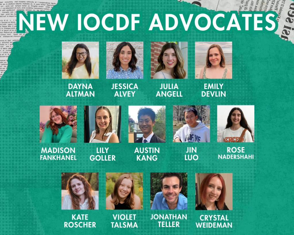
The IOCDF is thrilled to announce our newest cohort of Advocate volunteers! We’re welcoming 13 incredible new Advocates to our program, bringing our total to 64 dedicated volunteers working together to create meaningful change for the OCD and related disorders community.
These passionate individuals join us from bustling cities and quiet rural towns across the United States and around the world. From California to Massachusetts, and from countries including Canada and Ireland, this mix of local and global perspectives ensures we can reach and represent diverse communities everywhere.
The Power of Diverse Voices
Our newest cohort has a wide range of experiences and interests. They are passionate about addressing critical topics including:
This diversity of focus areas ensures that we can better represent and serve the full spectrum of our community’s needs.
Meet the Spring 2026 Advocates:
You can see the full list of IOCDF advocates at iocdf.org/advocate-program
Your Voice Matters Too
Inspired by our Advocates? You can make a difference! Here are ways to start advocating today:
Fuel Our Mission Through Fundraising
Turn your passion into action by launching a personal fundraiser. Whether for a birthday, a race, or a creative project, you can rally your friends and family to raise critical funds. Every dollar helps build a world where everyone affected by OCD can thrive. Start your fundraiser here or explore all ways to give back here.
Advocate for policy change
Your voice can shape laws that improve access to care and insurance coverage. The IOCDF Public Policy Action Center makes it simple to find the latest bills and contact your elected officials with just a few clicks. True change starts here.
Join an IOCDF Special Interest Group
Connect with people who share your experiences or professional interests. IOCDF Special Interest Groups (SIGs) provide a platform for deeper discussion.
Whether you advocate on the national stage, share your story to fight stigma, or fundraise your way, every action creates a ripple effect of hope and understanding. Your journey, your voice, and your commitment are powerful tools.
Start today and help us build a world where everyone affected by OCD feels supported, seen, and empowered. Join a dedicated community committed to raising awareness.
Welcome again to our new IOCDF Advocates, we’re grateful to have you joining our mission!
The post Welcome to Our New IOCDF Advocates appeared first on International OCD Foundation.
On 12 June 2026, Mental Health Europe joined more than 140 organisations working across children’s rights, digital rights, families and mental health in a joint statement to European Commission President […]
The post Joint Statement: Potential new measures for child online safety in the European Union appeared first on Mental Health Europe.
Background: Digital meditation-based interventions (MBIs) reach vast global audiences with millions of active users, yet concerns persist about the frequency and nature of adverse experiences (ie, AExs) occurring during meditation training. Some researchers have argued that AExs are substantially underdetected and reflect iatrogenic harm caused by meditation (ie, adverse effects [AEfs]). Others contend that these experiences largely reflect common stressors that would be experienced without meditation. These competing perspectives underscore the need for further research, particularly in the context of digital MBIs, the most widely used form of meditation training. Objective: This study examined the prevalence, predictors, and subjective evaluations of AExs during a digital MBI and tested whether reported experiences may be caused by meditation practice via comparisons between meditation-exposed and nonexposed participants. Methods: Data were drawn from 2 trials of the Healthy Minds Program. Exploratory study 1 (n=315) consisted of a sample of distressed US undergraduate students to estimate the prevalence of AExs and identify baseline predictors. Preregistered confirmatory study 2 (n=594) sampled distressed US adults from all 50 states to replicate findings from study 1 and to examine participants’ subjective evaluations of AExs. Study 2 additionally compared AEx rates between participants who did and did not complete guided meditations to assess whether AExs could be caused by meditation exposure. Study 3 (n=87) used qualitative methods to analyze study 1 participants’ responses to an open-ended question regarding their strategies for coping with AExs. Results: In studies 1 and 2, 27.9% (88/315) and 10.1% (40/396) of participants, respectively, reported at least one AEx during the study period, with 6.7% (21/315) and 3% (12/396) reporting functional impairment, largely aligning with previous research. Critically, in study 2, rates of AExs did not significantly differ between participants who did and did not complete guided meditations, suggesting that these experiences were not caused by meditation practice. Higher baseline depression, anxiety, loneliness, experiential avoidance, and perceived barriers to meditation predicted more frequent AExs. In studies 1 and 2, 89.8% (79/88) and 90% (36/40) of participants who reported AExs, respectively, indicated that they were glad to have learned to meditate. Qualitative analyses showed that participants used diverse coping strategies, often using skills learned through the Healthy Minds Program. Conclusions: AExs were relatively common but occurred at comparable rates among participants who did and did not meditate, challenging claims that such experiences were caused by meditation practice in distressed individuals. Although a small subset of participants reported some degree of functional impairment, most evaluated their AExs as tolerable and described their overall MBI experience as positive. Together, these findings highlight the importance of distinguishing AExs that likely reflect epiphenomena of preexisting distress or symptoms from iatrogenic harm attributable to MBIs. Trial Registration: Study 1: ClinicalTrials.gov NCT04741529; https://clinicaltrials.gov/study/NCT04741529; Study 2: ClinicalTrials.gov NCT06282523; https://clinicaltrials.gov/study/NCT06282523
<img src="https://jmir-production.s3.us-east-2.amazonaws.com/thumbs/09106e6bad7f3f15798304e0c00626ec" />
Background: The use of large language models (LLMs)–powered chatbots has reshaped how people seek information and advice, including for emotional and mental health support. While LLMs can offer scalable support, their ability to safely detect and respond to acute mental health crises—including suicidal ideation, self-harm, and violent thoughts—remains poorly understood. Progress is hampered by the absence of unified mental health crisis taxonomies, annotated benchmarks, and empirical evaluations grounded in clinical best practices. Objective: We addressed these gaps by introducing (1) a unified taxonomy of 6 clinically informed mental health crisis categories; (2) an evaluation dataset of over 2000 user inputs drawn from 12 publicly available conversational mental health datasets, classified into crisis categories; and (3) an expert-designed protocol for assessing response appropriateness. We also used LLMs to automatically identify crisis-indicative inputs and conducted an auditing study of 5 LLMs to evaluate the safety and appropriateness of their responses. Methods: We developed a taxonomy of mental health crisis categories informed by clinical experts and established literature. From over 239,000 mental health–related user inputs collected from 12 Hugging Face datasets, we curated 2252 examples (206 for validation, 2046 for testing) covering all taxonomy categories. We evaluated 3 LLMs on their ability to classify inputs into crisis categories, selecting the model with the strongest agreement with human annotators as the judge to label the test set. We then audited 5 LLMs on their ability to generate safe and appropriate responses to the 2046 test examples. Response quality was measured using a clinically informed 5-point Likert scale (1=harmful and 5=fully appropriate), relying on an LLM-as-a-judge validated against human expert feedback. Results: Several LLMs exhibited high consistency and generally reliable behavior when responding to explicit crisis disclosures, but significant risks remain. A nonnegligible proportion of responses was rated as inappropriate or harmful, particularly in the self-harm and suicidal ideation categories. Substantial performance differences were observed across models: gpt-5-nano and deepseek-v3.2-exp achieved very low harmful response rates, whereas gpt-4o-mini, Llama-4-Scout-17B-16E-Instruct, and grok-4-fast-non-reasoning generated markedly higher rates of unsafe outputs. All models exhibited systemic weaknesses, including poor handling of indirect or ambiguous risk signals, reliance on formulaic responses, and frequent misalignment with user context. Conclusions: These findings underscore the urgent need for enhanced safeguards, improved crisis detection, and context-aware interventions in LLM deployments and highlight the central role of alignment and safety engineering—beyond model scale or openness—in determining crisis response reliability. Our taxonomy, dataset, and evaluation framework lay the groundwork for ongoing research in artificial intelligence–driven mental health support, helping to minimize harm and protect vulnerable users.
<img src="https://jmir-production.s3.us-east-2.amazonaws.com/thumbs/6d38d493c2ebfaa303202d21ad5c3a7d" />
