For 18 months, the Trump administration has been accused by science advocates of shredding a social contract between academic researchers and the federal government that made the United States the world’s most powerful engine of discovery. Now it’s proposing a way to rebuild American science.
Taking inspiration from “Science — The Endless Frontier,” a 1945 manifesto that became the blueprint for the rise of American research universities, Trump’s science adviser, Michael Kratsios, this week put forth his version of such a report. Called “Science: A New Golden Age” and drafted by the White House Office of Science and Technology Policy, it calls for shifting hundreds of billions of research dollars from universities to industry, primarily tech and artificial intelligence companies.
The report acknowledges problems scientists have long attempted to grapple with, but advocates and scientists expressed concern that the report’s espoused goal of improving science is being undermined by other administration actions, chiefly the grievances expressed by President Trump and his aides against immigration, universities, and programs that they deem as promoting diversity, equity, and inclusion.
Conditions: Anorexia in Adolescence; Anorexia Nervosa; Anorexia Nervosa, Atypical; Anorexia Nervosa, Binge Eating/Purging Type; Anorexia Nervosa Restricting Type; Anorexia Nervosa With Significantly Low Body Weight; Body Composition Changes; Growth & Development; Malnutrition Severe; Malnutrition, Calorie
Sponsors: University of California, San Francisco; Eunice Kennedy Shriver National Institute of Child Health and Human Development (NICHD); Denver Health and Hospital Authority; University of Hawaii
Scientists at the Salk Institute and the Arc Institute, along with their collaborators, unveiled the first body-wide single-cell atlas of two major epigenetic systems: three-dimensional genome folding and DNA methylation, measured simultaneously in the same cells.
The atlas spans 86,689 cells from 16 human tissues, revealing 35 major cell types and 206 subtypes, and is freely available online. The work is part of the National Institutes of Health’s 4D Nucleome (NIH 4DN) program, which aims to understand how the genome is organized in space and time to regulate gene expression in health and disease.
Because the two epigenetic layers were measured together, the researchers could compare what each layer says about a cell’s identity. And while often the two pictures agree, they found that sometimes they do not.
The Human Genome Project, completed in 2003, produced a linear read of the three billion DNA letters in the human body. But the letters alone don’t explain how a single genome produces hundreds of different cell types. That information lives in the epigenome in the form of chemical modifications and structural folds layered on top of the DNA sequence, where they can switch genes “on” and “off” in patterns specific to each cell type.
Caption: Salk scientists Jingtian Zhou (left), Jesse Dixon (center), and Joseph Ecker (right) profiled 86,689 cells across 16 human tissues, linking cell-type-specific epigenetic features to disease risk and revealing that a cell’s 3D genome and DNA methylation don’t always tell the same story. [Salk Institute]
Two of the most consequential epigenetic features are 1) DNA methylation, where small chemical groups called methyl groups are attached to specific DNA bases, and 2) 3D genome organization, where intricate loops, folds, and compartments bring distant stretches of DNA into contact. Both influence gene expression, but they had never been measured together in single cells across the human body.
“There has been an appreciation for trying to understand, at the individual cell level, how the genome is organized, so that we can get a better idea of how genetic variants impact disease,” said co-corresponding author Joseph Ecker, PhD, a professor and Salk International Council Chair in Genetics at Salk and a Howard Hughes Medical Institute investigator. “Some cell types may be more vulnerable than others to genetic variants, because the genome is organized differently in different cell types—and whether a variant matters can depend on that organization.”
Why is noncoding DNA relevant in disease?
Most disease-associated genetic variants fall in the noncoding regions of the genome. That has made it difficult to figure out how a variant contributes to disease, which cell type it acts in, and what gene it ultimately affects.
The new atlas identifies more than 1.36 million differentially methylated regions and 283,606 differential chromatin loops across the human body’s cell types, using tissues from the heart, brain, lungs, stomach, skin, and more. When the researchers overlaid genetic variants known to raise disease risk, specific pairings emerged like variants for blood-glucose regulation concentrated in endocrine cells, atrial fibrillation variants in heart muscle cells, balding variants in skin fibroblasts, and bipolar disorder and schizophrenia variants in excitatory and inhibitory neurons.
“A lot of the genetic variation that predisposes someone to disease is in noncoding parts of the genome,” said co-corresponding author Jesse Dixon, MD, PhD, associate professor and Helen McLoraine Developmental Chair at Salk. “By adding in the 3D genome aspect, we can potentially bridge that gap—connecting noncoding variations with the genes they affect in specific cells and tissues.”
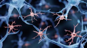
Microglia, illustration. Researchers from the New York Genome Center and Columbia University used the atlas’ cross-tissue methylation data to show that a substantial fraction of the brain’s resident immune cells (microglia) are replaced by cells resembling blood monocytes between roughly ages 50 and 75. The finding challenges the long-held view that microglia persist from embryonic development throughout the life span. [Artur Plawgo/Getty Images]
What happens when two epigenetic lenses disagree?
One of the study’s most surprising findings is that DNA methylation and 3D genome structure don’t always tell the same story about a cell. In skeletal muscle, the team found fibers that look like mature, differentiated muscle cells by their 3D genome folding, but still carry the methylation signature of muscle stem cells. The reverse almost never happens. The most plausible explanation, they explained, is that these cells are caught mid-differentiation, with 3D architecture updating first and methylation catching up.
Similar mismatches appeared in Schwann cells of the peripheral nervous system and in placental trophoblasts. The pattern suggests that different epigenetic features update on different time scales during cell state transitions—a finding that could reshape how researchers define “cell type” in adult tissues and how they track cells moving between states in disease.
The atlas also revises a long-standing assumption about “non-CG methylation,” an unusual form of methylation previously thought to be largely confined to brain cells and stem cells. The study shows that it carries cell-identity information across many human tissues, including muscle, pancreas, and immune cell types, at lower but biologically meaningful levels.
“The inconsistency between modalities may be further used to determine what cell populations are switching between each other in adult tissues and diseases, which could, for example, expand our understanding of cancer cell dynamics,” said co-first and co-corresponding author Jingtian Zhou, PhD, a former graduate researcher in Ecker’s lab who now leads his own lab at the Arc Institute.
A public resource for scientists and artificial intelligence
To make the atlas broadly usable, the team built an interactive web browser that lets researchers visualize DNA methylation and 3D chromatin contacts across every tissue, cell type, and subtype in the study. The underlying data, including 195 billion methylation measurements and 18 billion chromatin contacts, are freely available.
The resource arrives as artificial intelligence tools are increasingly used to predict the functional impact of genetic variants. Atlases like this one can provide the labeled, cell-type-resolved training data that models need to make accurate predictions—a bottleneck that has historically limited the field.
For example, in a companion paper in the same issue of Science, a study led by Bing Ren, PhD, from the New York Genome Center and Columbia University used the atlas’ cross-tissue methylation data to show that a substantial fraction of the brain’s resident immune cells, called microglia, are replaced by cells resembling blood monocytes between roughly ages 50 and 75. The finding challenges the long-held view that microglia persist from embryonic development throughout the life span.
“DNA methylation patterns are specific to each cell type and analogous to a cellular barcode,” said Ren, who also co-authored the Salk-led study. “The comprehensive cross-tissue DNA methylation atlases show that the aging microglia in the human hippocampus more closely match the monocytes from peripheral blood than microglia from young adults, providing a crucial clue for the biological identity of these cells.”
The NIH 4D Nucleome consortium, of which this study is a part, aims to extend this kind of mapping into the fourth dimension: time. A 4D understanding of the genome—how its structure and chemistry change as cells develop, age, and respond to disease—remains a major goal, and the cross-tissue atlas provides reference scaffolding that future time-course studies will build on.
Along with scientists from the Salk Institute and Arc Institute, investigators from the University of California, San Diego, Columbia University, New York Genome Center, University of California, Los Angeles, Harvard, Henan University in China, Vanderbilt University, Stanford University, Broad Institute, University of Sheffield in the U.K., Yale, University of Florida, University of Freiburg in Germany, University of Graz in Austria, and Nanchang University in China; and Chongyuan Luo also contributed to the Science paper.
Designing and developing a new medicine is an expensive, failure-prone scientific challenge. A new drug can take many years to develop, at the cost of a significant investment. And even then, most possible candidates never reach the patient. For biologic medicines, therapies made from engineered proteins rather than synthetic chemistry (which are often used to treat conditions across most major acute and chronic diseases), the complexity is even greater.
Scientists explore vast quantities of possible molecules, looking for the rare few that will bind to the right target, remain stable in the human body, and be manufacturable at scale. Today, AI is speeding up these processes and has quickly become a core part of the infrastructure in pharmaceutical R&D.
AI-assisted design is a growing part of how biologic drug candidates are developed, and companies like AstraZeneca are actively building its engineering teams to push this further. “Everything we do, whether it’s design, make, test, or analyze, is now computationally enhanced,” says Puja Sapra, senior vice president and head of R&D biologics engineering and oncology targeted discovery at AstraZeneca. “The cycle times are getting shorter while productivity and innovation increase.”
Sapra explains that AstraZeneca’s approach follows a build-measure-learn loop. AI generates or prioritizes candidate molecules computationally, predicting which designs are most likely to succeed. Scientists then focus lab resources only on the top-ranked candidates. This leads to a tighter feedback cycle with fewer dead ends, faster iteration, and the ability to go after disease targets that were previously considered untreatable by medicine. Because the number of possible molecular combinations far exceeds what any human team can systematically explore, using AI to narrow and refine the options for testing has become a major focus in biologics drug design.
Navigating complex drug design problems
Beyond accelerating timelines, AI is also being applied to the discovery of entirely new classes of medicines. Traditional biologics typically target one disease pathway. The next generation of drugs can hit multiple targets simultaneously or precisely deliver therapeutic payloads to specific cells. Achieving this requires optimization across many variables at once. Looking ahead AI-driven models could help design these increasingly complex, multi-specific biologics, explains Puja Sapra. “For example,” she continues, “such models could help identify which two or three targets to prioritize based on the underlying biology, then optimize across multiple parameters to balance a molecule’s potency, stability, manufacturability, and safety.” “Drugging the undruggable is becoming a reality,” Sapra says. “These technologies will eventually enable us to develop medicines against targets once thought impossible to reach. The potential for benefit to patients is remarkable.”
The data moat
McKinsey estimates that generative AI, combined with other computational tools, could cut drug discovery timelines by as much as 50%. But every AI model is only as good as its training data. In drug discovery, that means ample quantities of high-quality biological data. Experiments can provide a rich source of such data. Whether they succeed or fail, each experiment generates a signal about what does and does not work.
“Data is our differentiator,” says Sapra, explaining how the company’s datasets are proprietary and multimodal and include molecular structures, binding measurements, safety profiles, and manufacturing outcomes. “We’ve built an intentionally diverse portfolio across multiple disease areas and drug types. All of that data empowers us to fine-tune frontier AI models with richer, more representative training sets.” She continues, “Further, we have invested in deep screening technologies to generate additional datasets required in volume to constantly refine and validate our models.”
Building an autonomous discovery engine
To bring all of that data together in one place, AstraZeneca is building what it calls a “lab of the future” facility in Kendall Square, Cambridge, Massachusetts where AI and robotic automation will be able to form a continuous, closed-loop discovery system. “Where a self-driving car uses sensors and models to navigate its environment, this system uses AI to make predictions, robotic systems to execute experiments, and instruments to generate data,” explains Sapra. That data feeds directly back into the models, accelerating each subsequent cycle.
“Throughout, scientists will remain central to the process, providing the oversight, judgement, and strategic direction that ensure outputs are explainable, tolerable, and directed toward potential patient benefit,” she adds.
Eventually, automated high-throughput systems will be able to make and evaluate thousands of molecular interactions on a weekly basis. “This will generate AI-ready data at a scale that traditional workflows cannot match,” Sapra says. “Robotic sample handling, automated quality checks, and integrated data pipelines also have the potential to help accelerate early drug development timelines significantly.”
The next frontier: Generating medicines from scratch
Ultimately, Sapra says, the end-state vision for AI in biologic drug discovery is what the field calls “de novo” design. For this, the goal is for AI to generate entirely new protein sequences that precisely fit the desired drug properties. This includes designing the structure, predicting safety, how it will behave in the body and how to make it manufacturable.
“The field is making great progress toward a completely AI-generated biologic, designed from scratch all the way to a clinical candidate,” Sapra says. “As we continue to leverage frontier models and fine-tune them with the right datasets, we bring ourselves closer to this reality. I believe it will come. It’s a matter of time.”
Several key elements are needed to reach this point, however. First is richer and more standardized training data across the industry. Second, robust evaluation benchmarks for AI-generated candidates. And third, teams that know how to work at the intersection of machine learning and biology. Of all the prerequisites, however, safety prediction may be the most consequential, and perhaps the least discussed, Sapra says.
“One of the hardest problems in de novo design is predicting whether a computationally generated molecule will be safe in the human body,” Sapra explains. AstraZeneca is tackling this with what amounts to virtual clinical trials. These are advanced cell systems and micro-scale organ models that function as physical testbeds, paired with AI that learns from their outputs.
“These systems have the potential to generate enhanced biological signals without traditional testing bottlenecks, and they’re a critical missing piece in closing the loop between AI-generated designs and clinical-ready candidates,” Sapra adds.
A shift currently underway is the move toward agentic AI systems that can simultaneously generate molecule candidates and predict how efficacious and safe they are likely to be. These autonomous workflows can connect disease-level insights directly to molecule design, bridging what were previously separate data silos. “The complexity of the biology goes hand-in-hand with the design of the molecule,” summarizes Sapra.
Human talent unlocks AI potential
The transformation underway in biologics is not just about technology. “With more autonomous systems, human oversight remains at the heart of this approach—ensuring explainable and ethical AI for the benefit of patients,” says Sapra.
For scientists, working with AI is a collaborative process. “Scientists will work hand-in-hand with these model systems,” she says. “There will be a world where models will design molecules, then scientists will work with the systems to test those molecules and put all that data together.” Through this process of human checks, balances, and judgement calls, the models will evolve and constantly improve, ultimately with potential to benefit patients.
For engineers, designing and building effective systems ready for human-AI collaboration will mean ensuring high levels of model transparency and explainability. According to Sapra, AstraZeneca’s engineering teams include data scientists, automation specialists, and AI engineers, who are developing systems that act as “thinking partners” rather than black boxes. “Engineers are designing systems that generate, validate, and learn at speed. And the problems are genuinely hard: Multimodal data fusion, closed-loop optimization, uncertainty quantification, and interpretability at the point of clinical decision-making,” she adds.
In taking on such technically demanding challenges, engineers and scientists have the opportunity to contribute to the research and development of potentially life-changing treatments for many diseases, says Sapra. “The biologic medicines we can develop today, and those we’ll design tomorrow, depend on combining world-class AI and engineering talent with deep scientific expertise.”
This article has been initiated and funded by AstraZeneca. Z4-85058, July 2026.
This content was produced by Insights, the custom content arm of MIT Technology Review. It was not written by MIT Technology Review’s editorial staff. It was researched, designed, and written by human writers, editors, analysts, and illustrators. This includes the writing of surveys and collection of data for surveys. AI tools that may have been used were limited to secondary production processes that passed thorough human review.
Scientists have created the most detailed map to date of single-cell gene regulation in heart failure. Published today in Science, the study integrates multiple layers of genomics data to identify new therapeutic targets for the world’s leading cause of death.
“Our goal is to use this atlas to discover targets that we can act on therapeutically,” said Neil C. Chi, MD, PhD, professor of medicine at UC San Diego School of Medicine. “Right now, one of the biggest limitations in cardiology is not the lack of tools, but the lack of targets. This kind of data changes that.”
While previous genetic studies have uncovered many genetic changes linked to heart failure, more than 85% of them were found in noncoding DNA regions, making it difficult to understand how they contribute to the condition and develop targeted therapeutics. Because of this, treatment options for heart failure remain limited.
“What makes this study unique is the ability to integrate multiple layers of genome regulation in the same cells,” said Bing Ren, PhD, professor emeritus of cellular and molecular medicine at UC San Diego School of Medicine and scientific director and CEO of the New York Genome Center. “These technologies allow us to look beyond which genes are active to understand how the genome is organized and controlled, revealing regulatory elements and interactions that were previously inaccessible.”
The researchers analyzed more than 750,000 individual heart cells from 36 participants with and without heart failure. Results revealed that heart failure is associated with major shifts in cell composition, with a reduction in the number of cardiomyocytes and an increase of fibroblasts and immune cells. In failing hearts, over 10,000 genes showed altered expression patterns and more than 50,000 DNA regions showed changes in chromatin states that altered the ability of proteins to interact with them.
Compared to other cell types, fibroblasts and cardiomyocytes showed the most extensive remodeling of their gene regulation networks. These cells also showed several distinct cell states as they progressed from healthy to diseased—in the case of fibroblasts, transforming into activated fibroblasts and myofibroblasts that contribute to scarring and fibrosis.
“These intermediate states are where the disease is actively unfolding,” said Chi. “If we can understand and target those transitions, we may be able to intervene earlier and more effectively.”
Integrating the single-cell atlas with genome-wide association data, the team showed that genetic changes are concentrated in specific regulatory regions active in certain cell types, particularly in cardiomyocytes. These findings suggest that targeting cardiomyocyte genes may be more effective than targeting genes found on other cell types when treating heart disease.
“This is a higher-order view of disease biology,” said Chi. “Instead of just asking which genes are turned on or off, we’re now understanding how their regulation is controlled across the genome—and that’s where most disease risk actually resides. By connecting genetic risk, gene regulation and cell-specific disease processes, this study provides a blueprint for precision therapies in heart failure. It opens the door to targeting the right mechanisms in the right cells at the right time.”
New data from a National Institutes of Health-funded study shows that midlife, the immune cell landscape of the hippocampus, undergoes substantial remodeling. It points to a potential mechanism by which aging may contribute to the chronic neuroinflammation commonly seen in neurodegenerative disease. Details are published in a new Science paper titled “Epigenetic and 3D genome reprogramming during the aging of human hippocampus.”
The work was done by a collaborative team of scientists from the University of California, San Diego, the New York Genome Center, and the University of California, Irvine. According to the paper, the scientists analyzed postmortem hippocampal tissue from 40 neurologically healthy adults aged 20 to 95 years old.
Digging into the details, the scientists used traditional measures of gene expression alongside more advanced techniques to analyze the genome’s 3D architecture and epigenome. “Gene expression tells us what a cell is doing today, but epigenetic signatures preserve information about where a cell came from,” said Nathan Zemke, PhD, director of single-cell genomics at the UC San Diego Center for Epigenomics and first author on the study. “By combining these approaches, we uncovered a major shift in the identity and lineage of immune cells in the aging human brain’s immune cells that gene expression data alone would not have revealed.”
They found that the brain’s primary immune cells progressively decline from age 50 to 75 years of age, and are replaced by cells with elevated inflammatory signatures and other features that resemble the characteristics of peripheral blood-derived immune cells. It raises questions as to whether microglia, which emerge during embryonic development, may not renew throughout the human lifespan as previously thought. The data also showed that cells that typically maintain the protective blood-brain barrier deteriorated with age. And across many brain cell types, aging accompanied a widespread and coordinated disruption of genome architecture.
“The progressive structural disruptions were closely linked to shifts in gene regulation and cell identity, potentially revealing a fundamental feature of aging in the human brain,” said Bing Ren, PhD, scientific director and CEO of the New York Genome Center, and professor of genetics and development at Columbia University. Ren is also a corresponding author on the study,
Future studies will investigate the mechanisms driving the loss of resident microglia and determine whether the newly identified immune-cell transition contributes directly to Alzheimer’s disease and other age-related neurological disorders. Insights from the current study as well as others could provide new opportunities to develop therapies that help to preserve brain function and reduce vulnerability to neurodegenerative disease.
When it comes to organ donation, time is everything. As soon as an organ has been carefully removed from a donor’s body, it starts to deteriorate. Surgeons have a matter of hours to get it into a recipient. Leave it too long and the organ will become unusable.
In most cases, organs will be kept on ice during that time, at around 4 °C (39 °F). They cannot be frozen—in previous attempts, ice has formed, causing all kinds of damage.
Matthew Powell Palm at Texas A&M University and his colleagues have an alternative solution—a device that allows organs to be cooled to -4 °C (25 °F) without forming any ice.
Now, in new research with pig organs, his team has shown that kidneys, at least, can be supercooled and preserved in the device for days. Once rewarmed, the organs have been successfully transplanted into animals, and they seem to do better than organs kept on ice.
The work represents “a landmark achievement,” says Kevin Myer, president and CEO of LifeGift, an organ procurement organization based in Texas, who was not involved in the research.
Cooling organs
Powell Palm hopes this approach could ultimately help ease the organ shortage crisis. Today, there are more than 104,000 people waiting for a kidney transplant in the US alone. It is estimated that 17 people die every day in the US while waiting for a transplant. That’s partly due to a lack of donated kidneys, but it’s also because many of those that are available never make it to a recipient. In some years, around one in three donated kidneys end up being discarded, often because they end up too degraded to use by the time they reach a recipient. Kidneys can be stored on ice for around 24 hours or placed in devices that aim to mimic the conditions of the body, also for up to around 24 hours. That’s not always long enough to find a suitable recipient and transport the organ, says Myer.
Scientists around the world have been working on ways to store organs for longer by cooling them to even chillier temperatures. Cooling an organ slows its metabolism—the colder you go, the greater the effect, and the longer you can store it.
We’ve long been able to successfully cryopreserve eggs, sperm, and embryos, but it’s much harder to freeze large organs. Teams have been exploring various temperatures and cryoprotectants (chemicals that essentially work like antifreeze), but so far no one has been able to freeze human organs for transplantation.
As a thermodynamicist, Powell Palm explored another approach. By keeping an organ submerged at a constant pressure, it should be possible to prevent the formation of ice at temperatures a little below 0 °C, without the need for cryoprotectants (which might have side effects and would need to be approved before being used in human transplants).
To test this theory, Powell Palm and his colleagues have created a device that does just that. The device itself is essentially a hermetically sealed chamber with a transparent lid. At its base is a device that monitors the organ’s temperature and checks for the formation of ice. Organs are submerged in a solution that is already commonly used to preserve them for transplant. “I always describe this as low-tech high science,” says Powell Palm. “A lot of work has gone into understanding the … kinetics at play in this system, but ultimately … it’s quite simple.”
Supercooled kidneys
To test their device, Powell Palm and his colleagues first removed single kidneys from pigs. The organs were flushed with the same commonly used solution to remove the blood, just as transplant organs are. The team then kept some kidneys on ice for either two hours or 24 hours, to mimic standard conditions used in human transplantation. They also put some of the removed kidneys in their device for 24, 48, or 72 hours.
The stored kidneys were then each transplanted back into the original donor pigs. Each pig’s second kidney was removed in the same procedure, leaving each animal with only the kidney that had been stored, and reimplanted.
Once the 24-hour supercooled kidneys were transplanted, they immediately began producing urine—a key indication that they were working. The team members also measured other markers of kidney function and found that the organs appeared to be working normally within about 10 days of being transplanted.
A kidney that was supercooled for 72 hours recovers once it is transplanted back into a pig.
COURTESY RONALD SELLERS, POWELL-PALM LAB, TEXAS A&M UNIVERSITY
That’s slower than kidneys stored on ice for two hours but much quicker than kidneys kept on ice for 24 hours, says Powell Palm.
The organs that were kept supercooled for 48 and 72 hours performed similarly, he says. “Even at three days—triple the clinical standard—we’re getting recovery that is faster than … [what has been] the gold standard for the last three decades,” he says. “So we’re really, really pumped about this.”
“It is impressive,” says Heidi Yeh, a transplant surgeon at Mass General Brigham for Children, who also researches organ preservation technologies. “Often kidneys that have been stored for 48 hours [in other studies] take a week or two before they start working again.”
Organs that grow
The supercooled organs seem to work well in the long term, too. Over a 30-day period, the pigs grew by around 30%—and the kidneys grew with them, almost doubling in size to compensate for both the pigs’ growth and the lack of a second kidney. The team monitored one of the pigs for 200 days before removing and analyzing its kidney. Even at that point the organ looked healthy, says Powell Palm. He and his colleagues presented the findings at the American Transplant Congress in Boston last month.
Earlier this year, researchers in Canada showed they could also cool pig kidneys to below-zero temperatures and transplant them into pigs. The team’s protocol included the use of a cryoprotectant, and organs were stored for up to 48 hours before being transplanted into pigs. Those organs survived for a week.
In supercooling organs for 72 hours and showing that they do well for 30 days or more, Powell Palm and his colleagues have broken new ground. “It’s the first time this has ever been reported in history,” he says.
Those extra hours could make all the difference, says Myer of LifeGift. The advance could give doctors more time to evaluate the kidneys, match them to the most suitable donors, and physically get the organs to their intended recipients in time. It could enable international donations and open up cheaper transport options, he adds. “Right now, with kidney transplantation the assumed limit is 18 to 24 hours,” he says. “If we can get up to 72 hours … that would change everything.”
Powell Palm and his colleagues think they may even be able to go beyond 72 hours. In preliminary studies, organs that had been stored for up to 120 hours appeared healthy, although those organs have not yet been transplanted.
And because the process doesn’t require any cryoprotective chemicals, the team members are hoping for an accelerated approval from the US Food and Drug Administration, which would allow them to test the device in human transplantations.
The storage device is simple and compact, so Powell Palm thinks it will be easy to transport. It hasn’t been tested for air travel yet, but it has been used to take supercooled kidneys across the US in the back of a Kia Sorento, he says: “From a stability perspective, we view this as an even higher bar.”
Powell Palm and his colleague Sebastian Giwa plan to launch a company dedicated to developing the technology, along with other protocols that “stop biological time,” in the coming months, he says.
This is today’s edition of The Download, our weekday newsletter that provides a daily dose of what’s going on in the world of technology.
The power line that could reshape New York’s grid is hitting snags
During a heat wave on July 3, New York State’s grid imported enough electricity from Canada to meet about 9% of its total demand that day.
Some of that power shuttled in on a 339-mile power line stretching from Quebec to Queens. It opened in May and is officially the longest underground transmission line in North America. It could provide up to 20% of New York City’s electricity demand, largely with abundant hydropower from Quebec.
One wrinkle: The line has been down for most of this month, and some experts are concerned about how drought will affect the power supply feeding it.
Still, the line could help shape the future of our grid, if it can overcome these sorts of snags. Read our story to understand how.
—Casey Crownhart
This story is from The Spark, our weekly climate tech newsletter. Sign up to receive it in your inbox every Wednesday.
The must-reads
I’ve combed the internet to find you today’s most fun/important/scary/fascinating stories about technology.
1 The US Treasury is threatening to sanction Chinese AI companies Treasury secretary Scott Bessent has accused Moonshot of improperly distilling Anthropic’s Fable model. (TechCrunch) + Nvidia’s Jensen Huang is arguing that America has nothing to fear from Chinese AI. (Axios) + Like it or not, Chinese models are now part of the global AI infrastructure. (Rest of World) + China’s AI models have Trump’s AI world at war with itself. (MIT Technology Review)
2 Why the OpenAI hack is the scariest AI mishap yet AI’s capabilities seem to be starting to outpace our current ability to control them. (The Economist $) + Hugging Face had to turn to a Chinese AI model to rescue it from the hack. (BI)
3 Visually impaired Europeans can now get an implant that restores sight And Americans may not have to wait long to receive it, too. (STAT) + This retina implant lets people with vision loss do a crossword puzzle. (MIT Technology Review)
4 A bellwether lawsuit suing Meta for social media addiction has been dropped There are, however, many more waiting in the wings. (NYT $)
5 Here’s how ICE gets its hands on Americans’ data As soon as you open a credit card or phone account, its agents can see where you live. (404 Media) + States are warring with the Trump administration over the right to see ICE agents’ faces. (Wired $)
6 We urgently need to grapple with AI’s environmental impact As the world warms, is the price we’re paying worth it? (The Verge) + We did the math on AI’s energy footprint. (MIT Technology Review)
7 Privacy issues with smart glasses need an industrywide fix That’s according to Samsung, which is unveiling glasses it developed with Google this fall. (Bloomberg $)
8 The US Army is begging soldiers to limit their AI use The token crisis comes for us all eventually, it seems. (Ars Technica)
9 Why does lettuce keep making Americans sick? It’s pretty simple: a lot of people eat it, and it doesn’t get cooked. (Wired $)
10 Pokemon Go is the perfect game to play this summer It’s fun, collaborative, and it gets you outdoors. (Guardian)
Quote of the day
“It went off and did this hack all by itself, as far as we can tell. This is the highest level of autonomy that we’ve seen in the use of a large language model for cyber operations.”
—Colin Shea-Blymyer, a cybersecurity research fellow at Georgetown University, tells NPR why the OpenAI hack on Hugging Face is so alarming.
One More Thing
KAGAN MACLEOD
Welcome to the dark side of crypto’s permissionless dream
Jean-Paul Thorbjornsen is a founder of THORChain, a blockchain through which users can swap one cryptocurrency for another and earn fees from making those swaps.
But is he responsible for what it’s used for? It’s a question that matters because in January last year, its users lost more than $200 million in cryptocurrency after THORChain transactions and accounts were frozen by an admin override, which users believed was not supposed to be possible given the decentralized structure. It’s also been used by North Korean hackers to move $1.2 billion of stolen ethereum.
A place for comfort, fun, and distraction to brighten up your day. (Got any ideas? Drop me a line.)
+ A musician developed an ingenious way to strum a guitar with an electric fan. + Ukraine’s tunnel of love is a leafy green corridor of romance that’s straight out of a fairy tale. + The driver of a giant banana has been pulled over 100s of times, but still won’t ditch his treasured ride. + Ever wonder which albums and songs truly stand the test of time? The Greatest Music tries to answer that via an algorithm that analyses hundreds of “best of” lists.
IntroductionDepression is among the leading causes of disability globally. Therefore, exploring the various non-medical treatment options for this condition is particularly important. The aim of the review was to assess the effect of art therapy on depressive symptoms.MethodsThe foundation of this review is a pre-planned, explorative, secondary analysis of a previously published umbrella review, encompassing the databases Cochrane Library, Embase, MEDLINE, CINAHL, ERIC, American Psychological Association PsycArticles, American Psychological Association PsycInfo, PSYNDEX, the German Clinical Trials Register, and ClinicalTrials.gov. Included were all randomized trials with any patient population receiving active visual art therapy. The outcome was depressive symptoms measured by depression assessment instruments. We followed the Preferred Reporting Items for Systematic Reviews and Meta-Analyses (PRISMA) guidelines and conducted a bias assessment using a modified Cochrane risk of bias tool. Data was pooled using a random-effects model and visualized in forest plots. A pooled standardized mean difference (SMD) with hedges g was calculated to measure the reduction of depressive symptoms.ResultsOf 3,100 identified reports we included 26 studies. Of these, 19 studies with 997 patients were eligible for inclusion in the meta-analysis. Overall, we found a standardized mean difference of 0.53 (95% CI: 0.30 to 0.76) for depressive symptoms, favoring the intervention group. Main sources of variation were different types of control groups, methodological quality, and patient populations.ConclusionOur results suggest that art therapy is associated with improved depressive symptoms. Therefore, art therapy should be accessible as complementary treatment for patients suffering from depressive symptoms.


![Caption: Salk scientists Jingtian Zhou (left), Jesse Dixon (center), and Joseph Ecker (right) profiled 86,689 cells across 16 human tissues, linking cell-type-specific epigenetic features to disease risk and revealing that a cell’s 3D genome and DNA methylation don’t always tell the same story. [Salk Institute]](https://www.genengnews.com/wp-content/uploads/2026/07/260723-pr-ecker-dixon-authors-232x300.jpg)