Part of the Hong Kong Genome Project, genomic analyses of more than 20,000 participants provide information on clinically relevant variants for the Chinese population and offer insights on the implementation of genomic medicine initiatives.
WASHINGTON — Over a recent breakfast, U.S. officials had a message for the German ambassador: pay more for pharmaceuticals.
The meeting, between U.S. Trade Representative Jamieson Greer, chief health department adviser Chris Klomp, and German Ambassador Jens Hanefeld, was part of a larger push from the Trump administration to get other countries to pay more for medications as the U.S. pays less, according to a person familiar with the meeting.
The U.S. officials discussed the possible use of tariffs under Section 301 — which grants the government authority to combat trade practices considered “unfair.” The move would be similar to the tactic of threatening new tariffs to get other countries to pay more for drugs. The ambassador agreed to review the matter with officials in Germany, but no deal was made, the person said.
The Justice Department sent a letter to Yale School of Medicine on Thursday, alleging it was illegally discriminating against applicants who are not Black or Hispanic, following a similar missive sent last week to the University of California, Los Angeles medical school.
A 2023 Supreme Court ruling prohibited the use of affirmative action in admission decisions, but the Trump administration accused the medical schools of continuing discriminatory practices despite that decision, citing differences in average test scores and GPAs between students of different racial groups over the past three admissions cycles.
At the ripe old age of 73, I take six drugs — four prescription and two over-the-counter pills — every day. I keep careful track of them.
But recently, when I received a refill, I noticed my blue pill was now yellow. I was startled, but this was not the first time my pills had morphed into different colors. A few months ago, my white and blue capsule had become white and purple, and before that, my pink tablet turned blue.
The evidence base for ADHD pharmacotherapy has answered one question more confidently than any other: whether medications are effective, on average, in reducing core ADHD symptoms. We know that several stimulant and non-stimulant treatments, including methylphenidate, amphetamines, atomoxetine, and guanfacine, improve symptoms at the patient-group level.1 What has remained harder to identify is where titration should stop: the point at which further dose escalation is unlikely to yield meaningful additional benefit and might instead worsen tolerability.
For years, artificial intelligence (AI) has been growing behind the scenes of our lives. Starting off as modifications of not‑so‑simple algorithms, early large language models could barely string a few words together, much like early vision systems that struggled to distinguish a lamppost from a cat in digital images. More recently AI has not just grown but proliferated—like Darwin’s finches in the Galapagos—into nearly every niche available in the digital world.
AI has infiltrated into daily life personally and professionally for many, and while modern healthcare has historically been hesitant to adapt to new technologies, Raghav Mani, director of Digital Health at Nvidia, pointed out that healthcare is adopting AI at three times the rate of other industries. Clearly, there is a lot to discuss, which is why The New York Academy of Sciences and the Windreich Department of Artificial Intelligence and Human Health at the Icahn School of Medicine at Mount Sinai co-hosted the 3rd annual “New Wave of AI in Healthcare,” a two-day symposium on May 12 and 13 with the goal of opening discourse between researchers, clinicians, industry leaders and other interested parties on all topics related to AI and healthcare.
Day one
The first day opened with a lightning round of welcome remarks from organizers expressing their personal experience with AI in healthcare research and practice. While some, like Nicholas Dirks, PhD, president and CEO of The New York Academy of Sciences shared concerns about how to maintain human involvement in AI use, he also expressed awe stating that “The pace of progress is breathtaking.”
Others were more practical in their assessments. Lisa Stump, chief digital information officer at Mount Sinai Health System asserted, “The future is not something we enter, it’s something we create.” Similarly, Brendan G. Carr, MD, CEO, Mount Sinai Health System, described AI as a “new partner” to aid clinicians in synthesizing the vast and growing clinical data. Girish N. Nadkarni, MD, a nephrologist and practicing clinician at Icahn School of Medicine at Mount Sinai summarized the whole event before the first talk even began: “The real question is not IF AI will transform healthcare, but HOW.”
The keynote presentation leading day one’s discussions endeavored to answer that very question. With his talk entitled, “Harnessing the power of Platform Thinking to Transform Healthcare,” John Halamka, MD, president of the Mayo Clinic Platform, spent 30 minutes exploring the power of data while questioning how AI is and should be used to analyze the varied data currently available, but cautioned that this is no simple task when considering the sources of data and potential restrictions on data use. He spoke about practical applications of AI data analysis that have and can be done, including in drug discovery. He also pointed out that AI can fill gaps in the healthcare workforce.
The day continued with four talks exploring different aspects of AI model use in healthcare. Marina Sirota, PhD, professor at the University of California, San Francisco spoke about how clinical data can be used for predictive medicine. Others, including Mani and Jonathan Carlson, PhD, vice president and managing director of Microsoft Heath Futures, discussed how AI agents and models can be used as part of hospital and clinician toolkits at multiple levels—not just as data analysis engines, but also to aid in synthesizing patient data and diagnostic support. Rounding out the discussion, Azra Bihorac, MD, senior associate dean for research at the University of Florida described how AI models need to be validated just like any other tool. She also pointed out that while AI is continuously improving in its ability to assess problems and suggest the next best course of action, human input is vital for collaborative success.
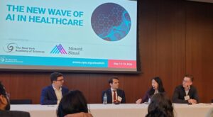
Panel discussion moderated by Robert Freeman, DNP. Panelists from left to right: Pierre Elias, MD, Karen Wong, MD and Alexander Fedotov, PhD
The final talks for day one focused on how AI can be used directly with patient care situations. Following their individual talks on how AI can be integrated into electronic health records (EHR), combining models to develop new insights, or reimagining diagnosis ability to improve diagnostic equity, the final three speakers engaged in a dynamic, and sometimes heated panel discussion. Karen Wong, MD, a physician at Epic, Alexander Fedotov, PhD, director of AI digital precision health at AstraZeneca and Pierre Elias, MD, assistant professor at Columbia University Irving Medical Center each shared their thoughts on how AI will be used in the near future. While they were all in agreement that AI cannot replace clinicians, they also recognized that AI will be a disruptive force, but it’s up to clinicians to take responsibility to use the technology as appropriate but to rely on their intuition and judgement as trained professionals. When opining on the future of AI use in healthcare five years from now, Fedotov stated, “I would still want to see humans at the helm of all the decision maker processes.”
Day two
While the first day laid the foundations for AI use in healthcare spanning bench to bedside, the second day of the symposium included more discussion and criticism of AI on the logistic level.
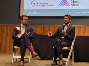
Fireside chat between Girish N. Nadkarni, MD and Dave A. Chokshi, MD
The day began with a keynote fireside chat between Nadkarni and Dave A. Chokshi, MD, a physician and professor at City University of New York, and former NYC health commissioner. He spoke about his leadership experiences, sharing many anecdotes of his time as a public health advocate and communicator during the COVID-19 pandemic. When questioned on the importance of communication considering the state of healthcare and declining trust of the public—especially with the increased use of AI, which has the potential of adding layers of feelings of abandonment, surveillance, and impersonalization—Chokshi pointed out that “It makes relationships even more important that we know then are.” He stressed that a his job, as a clinician, is to build trust with patients, and make sure that they return for care. While he envisions AI being transformative to healthcare in the next few years, he cautioned that listening and integrating feedback from front line users, clinical staff and patients, will be vital.
The morning continued with talks exploring AI’s use in research and learning in healthcare. Joshua C. Denny, MD, CEO of NIH All of Us Research, delivered a detailed summary of the progress and of the All of Us project. Despite recent funding concerns and cuts, the project scope remains on track, and researchers world-wide are utilizing the data derived from this project and how the project leads are working to establish parameters and modules for researchers to more easily implement AI in their data analysis. Andrew Gruen, PhD, standards lead at MLCommons, then spoke animatedly about the importance of establishing standards and benchmarks for AI use in researcher and healthcare settings. He spoke candidly on the need to not just train AI but to have external evaluation and validation of AI models.
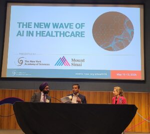
Panel discussion moderated by Girish N. Nadkarni, MD. From left to right: Karandeep Singh, MD, Girish N. Nadkarni, MD, and Vardit Ravitsky, PhD
The symposium concluded with multiple discussions on the interactions between AI and humans—not just as a tool, but by viewing the use of AI in the broader scale. Karandeep Singh, MD, executive director for health innovation at the University of California, San Diego explored various opinions of clincians and patients on the use of AI, while pointing out that the use of AI in healthcare settings should be thoughtfully considered before implantation. Meanwhile, Vardit Ravitsky, PhD, president and CEO of The Hastings Center for Bioethics, discussed the ethics behind AI use as a direct to patient setting, specifically as a patient-used chatbot. In a debate following their respective talks, the two delved deeply into the risks associated with AI use, both on the patient side with chatbots and with scribe technologies used by clinicians and patients. They often agreed on the need for transparency in AI usage, but specific AI applications, like uses of AI robots in the home to combat loneliness in the elderly resulted in disagreements.
The final talk presented by Tanzeem Choudhury, PhD, chief of health innovation at Cornell Tech, brought many previously discussed topics together. Her research explores how AI can be used in treatment of mental health, describing how AI can be used in multiple aspects of mental health therapy from recording physiological symptoms with wearables to using chatbots for various functions. She cautioned that while these tools may eventually be transformative, the current state of AI use in mental health is still growing.
The closing remarks by Alexander Charney, MD, PhD, professor at Icahn School of Medicine at Mount Sinai summarized the event well. He shared that throughout the symposium he imagined what clinicians and researchers from 100 years ago and from 100 years in the future would think about the current state of healthcare and about the challenges being faced now with how to incorporate AI. He said, “We aren’t the first group of human beings to deal with powerful technology and figuring out how we’re going to use it to change society.” He hopes that the people from the past would see that we understand and respect the past and learn from it being rigorous in our research and testing, while the people from the future will look on us with pride at our fearless and tenacity in the face of new technology. He hopes that both groups would see that we “tried to do the right thing.” He ended saying that he does see all of that here along with passion and coming together of everyone at the meeting.
<strong>Background:</strong> Tardive dyskinesia (TD) is a common, often underrecognized movement disorder resulting from long-term antipsychotic use, yet its detection in routine mental health care remains inconsistent despite the availability of structured rating scales. <strong>Objective:</strong> This study evaluated the performance of an artificial intelligence–powered, video-based model for detecting abnormal movements associated with TD using the Clinician’s Tardive Inventory (CTI) dataset. We compare automated assessments of videos from the CTI dataset with previously completed clinician-rated Abnormal Involuntary Movement Scale (AIMS) and CTI scores for the dataset’s videos to determine the model’s reliability and the accuracy of its assessment conclusions relative to expert raters. <strong>Methods:</strong> In total, 69 videos with corresponding AIMS and CTI ratings were analyzed using the visual transformer algorithm model called TDtect reported previously. The dataset included single-video assessments per participant, with varied instructions and movement types. The relationship between automated predictions and clinician ratings was assessed using Pearson correlation, and predictive accuracy was evaluated using area under the curve (AUC) metrics. <strong>Results:</strong> The model showed a strong correlation with AIMS total scores (<i>r</i>=0.717) and high diagnostic accuracy (AUC 0.854), which improved further at an optimized threshold (AUC 0.900). Performance differed across anatomical regions, with the tongue, lips, and jaw displaying the highest predictive reliability. Functional CTI components had weaker correlations (<i>r</i>=0.27-0.63), as expected due to the subjective nature of these measures. <strong>Conclusions:</strong> These findings provide preliminary evidence that an artificial intelligence–driven TD detection model can generalize across video protocols, suggesting potential for broader clinical applicability, although further validation is needed. Future refinements and fine-tuning are expected to enhance accuracy, particularly in predicting functional impact.
Researchers have found that a retina that is aging faster than usual can indicate a lower bone density and an increased risk of fractures due to osteoporosis. Published today in PLOS Digital Health, these findings set the basis for a novel diagnostic method for a condition that remains underdiagnosed due to a lack of accessible screening tools.
“Osteoporosis is a common condition that weakens bones and raises the risk of fractures, especially in older adults,” write the study authors, who were led by Ching-Yu Cheng, MD, PhD, professor at the Duke-NUS Medical School and director of the Singapore Epidemiology of Eye Diseases (SEED) program. “However, many individuals are not diagnosed until after a fracture occurs, in part because the standard diagnostic test, dual-energy X-ray absorptiometry (DEXA), is not always readily accessible.”
A DEXA scan uses very low levels of X-ray radiation to measure a patient’s bone density—a major indicator of osteoporosis and fracture risk. However, it is a costly procedure requiring specialized equipment, and therefore typically only recommended for high-risk individuals with suspected fractures on X-rays or patients on long-term steroid therapy. This limits early detection in the broader population, with many people being diagnosed with osteoporosis only after experiencing a fracture.
Worldwide, nearly 20% of the population is affected by osteoporosis. If left untreated, this condition increases the risk of major fractures, which can be life-threatening and represent a large economic burden for healthcare providers. This drives an urgent need for accessible and non-invasive screening methods that can replace traditional DEXA scans.
Cheng’s team investigated whether images from a patient’s retina could help identify those at a higher risk of developing osteoporosis. This idea stemmed from previous research indicating that the retina can reflect the body’s overall biological aging.
The researchers developed a deep learning algorithm, known as RetiAGE, which calculates the probability of a person being older than 65 years based on images from their retina. They then investigated whether there was an association between RetiAGE results and bone mineral density (BMD) scores, as well as osteoporotic and hip fracture risk scores calculated using the fracture assessment tool (FRAX).
Retinal images and DEXA measurements were obtained from 1,965 participants in the PopulatION HEalth and Eye Disease PRofilE in Elderly Singaporeans (PIONEER) study. In this patient population, older RetiAGE scores were linked to lower BMD scores and an increased risk of major osteoporotic and hip fractures.
The ability of RetiAGE to predict the onset of osteoporosis was then evaluated in another 43,938 participants from a prospective UK Biobank cohort with retinal photographs and no osteoporosis at the time of taking these images. Higher RetiAGE scores, indicating accelerated retinal biological aging, were able to predict future osteoporosis onset even when adjusting for common osteoporosis risk factors as well as female-specific risks such as menopause and hormone replacement therapy.
“These findings suggest that retinal biological aging may reflect broader aging processes related to skeletal health,” state the researchers. “Retinal imaging may therefore provide a simple, non-invasive, and accessible way to support opportunistic screening for osteoporosis risk.”
A next-generation device that detects signs of stress could have wide-ranging applications, from investigating sleep disorders to detecting signs of sepsis.
The polygraph detector, described in Science Advances, is worn on the chest and can even sense when a person is lying.
It allows psychophysiological states to be continuously monitored through a combination of multimodal sensing and wireless data transmission.
The gadget offers an alternative to current approaches such as such as polygraphy and polysomnography (PSG), which involve cumbersome wired sensors that limit their practicality.
“By uncovering mechanistic links between autonomic imbalance, stress reactivity, and health outcomes, these devices have the potential to transform diagnostic workflows, optimize educational programs, and enable personalized therapeutic monitoring across stress medicine, pediatrics, and behavioral health,” reported Sun Hong Kim, PhD, from the University of Seoul in South Korea, and co-workers.
Subtle physiological variations in cardiac, respiratory, electrodermal, and thermal activity often serve as indicators of compromised health or heightened stress responses.
These can be reflected in many scenarios, from pediatric sleep disorders that disrupt neurodevelopment to the psychological strain experienced in high-stakes clinical settings or during polygraph examinations.
Accurate monitoring of psychophysiological states is therefore essential for understanding how stress and autonomic dysfunction manifest across a wide spectrum of medical conditions.
However, most existing devices monitor only one or two parameters or rely on electrochemical sensors that detect sweat biomarkers, thereby failing to reflect the complex and dynamic interplay between multiple physiological systems.
Wearable polygraph device in the palm of a hand for scale. [John A. Rogers/Northwestern University]
Kim and co-workers therefore designed a single platform to enable comprehensive assessment of autonomic and stress-related physiology in real time.
The device continuously measures changes in heartbeat, skin temperature, and breathing, which are then converted using machine learning into measures of psychological strain.
The device had high fidelity with gold standard systems in quantifying the complex psychological stress induced by polygraph interviews and complex cognitive load tasks as well as the physical stress caused by repeatedly putting a hand in an iced water.
During overnight monitoring of children, it reliably identified arousals, hypopnea, and apnea while revealing disease-specific autonomic signatures among infants with Down syndrome.
Real-world deployment during emergency simulation training showed that multimodal stress signatures correlate inversely with performance, reflecting its value for medical education.
Machine learning analyses across all studies confirmed that multimodal features outperformed single-signal approaches in detecting stress and clinical events with high sensitivity and specificity.
“A particularly notable contribution lies in pediatric sleep medicine,” the authors noted.
“Simultaneous comparison with PSG confirms the ability to detect arousals, hypopnea, and apnea while also providing mechanistic insights into autonomic regulation.
“In infants with Down syndrome, multimodal analysis reveals attenuated sympathetic responsiveness and parasympathetic dominance, consistent with known vulnerabilities in airway patency and autonomic control.
“Such disease-specific autonomic signatures may serve as valuable biomarkers for risk stratification, early diagnosis, and targeted intervention in neurodevelopmental disorders.”