

Synthetic Organizers Aid Creation of Reproducible Kidney Organoids from Stem Cells

University of Southern California (USC) researchers have paired a biological discovery with an engineering feat to create more faithful, reproducible kidney organoid structures, grown from human pluripotent stem cells (hPSCs). By mapping the developing human kidney, the scientists identified a previously unrecognized developmental axis that helps organize the kidney’s nephrons, which are their filtering units. The team then engineered Wnt-secreting “synthetic organizer” cells to recreate aspects of this developmental environment in organoids.

Their advance makes the organoids more reliable models for studying disease and evaluating potential therapies, while supporting long-term efforts to generate transplantable kidney tissue. “It is important that we’re starting to get good reproducibility from organoid models that can lead to robust preclinical models of cell function and disease to benefit patients,” said Nils Lindström, PhD, assistant professor of stem cell biology and regenerative medicine at the Keck School of Medicine of USC. Lindström is co-corresponding author of the team’s published report in Science, titled “Patterning human kidney organoids with synthetic Wnt-secreting organizers.” In their paper, the researchers reported, “Our findings link a spatial organizing geometry in the developing human kidney to controllable engineering in vitro.”

“Stem cell–derived organoids have emerged as systems for modeling organ development and generating complex tissue structures in vitro,” the authors wrote. Over the past decade, organoid work has relied on cells’ ability to self-organize into tissue-like structures, often in response to adding chemicals and proteins that act broadly in the whole organoid. “Although this capacity enables organoids to recapitulate many developmental programs, it limits experimental control over tissue architecture, often producing structures that vary between cultures and are difficult to engineer reproducibly,” the team continued. “Understanding how to impose spatial patterning in organoid systems is therefore an important challenge.”

In embryos, spatial patterning is often organized by localized signalling centers, known as developmental organizers. But how organizing geometry is controlled in the developing kidney, and whether it can be recreated in vitro, hasn’t been known.

For their reported study, the team combined spatial transcriptomics of the developing human kidney with synthetic engineering. “We mapped this organizing geometry in developing human kidneys and tested whether a minimal cue of localized WNT signaling could restore spatial control of nephron patterning in organoids,” they explained.

The project began by making tools to copy developmental signals. Postdoctoral researcher Fokion Glykofrydis, PhD, in the Morsut lab, engineered a “synthetic organizer” cell that secreted a Wnt protein that their spatial transcriptomics and other analyses indicated was involved in spatial patterning during kidney development. Graduate student Connor Fausto from the Lindström lab proposed an experiment to test how this Wnt-secreting cell would affect organoid nephrons.

The experiments revealed that the synthetic organizer enabled two key processes essential for building organs: controlling the identity of cells and influencing the shape of developing structures. The synthetic organizer serves as a localized and targeted source that secretes controllable amounts of specific Wnt proteins within the organoid itself. These are key signals that help shape the developing kidney. This creates a signaling environment much more similar to a naturally developing kidney and gives researchers a way to control where and how kidney structures form.

Co-corresponding author Leonardo Morsut, PhD, associate professor of stem cell biology and regenerative medicine, and biomedical engineering at the Keck School of Medicine and USC Viterbi School of Engineering, said, “With our approach, we are trying to control self-organization, and work with it as opposed to try to completely override it.”

Lindström expected Wnt to trigger nephrons to change their identity into cells capable of forming connections with the urine drainage system. What surprised him was that the nephrons also changed shape and elongated toward the source of the Wnt signal, which doesn’t happen when signals are delivered uniformly to the whole organoid. Compared with the developmental process seen in traditional kidney organoids, this elongation toward the Wnt source is more similar to what happens in a naturally developing kidney.

“A single, localized signal did two things at once. It changed what the cells became and physically pulled the tubules toward the source,” Lindström said. “You would not see that with a uniform chemical bath of signals.” Engineered WNT-secreting cellular organizers introduced into kidney organoids restored organizing geometry, the authors noted, “… biasing distal nephron differentiation and orienting nephron morphogenesis toward the signal source, which demonstrates that developmental signaling geometry can be reconstructed synthetically to control tissue patterning.”

The team identified a previously unrecognized axis, a direction along which the developing kidney organizes itself. Developmental biologists have long known about the nephron’s classic “proximal-distal (PD) axis,” which runs from its blood-filtering end to its urine-drainage end. The new axis is defined instead by how close each part of the nephron sits to the collecting duct, the tube system that drains urine and releases Wnt signals during development. Those signals tell the nephron what shape to take and which way to point.

“The study shows that there’s an undiscovered axis that sets up how a nephron looks and forms,” said Lindström. “It’s not every day that you find something new in human development at that level.”

Most kidney organoids contain only nephrons and lack the collecting duct that supplies this local Wnt signal, so they have no such axis and organize in a radially symmetrical pattern. By mapping how kidney cells respond to Wnt at specific locations in the developing kidney, the team recreated that environment in organoids with the synthetic organizer, producing structures that are both more developmentally faithful and more reproducible.

“Introducing tunable WNT-secreting synthetic organizers (SOs) in organoids restored canonical WNT responses, biased distal nephron differentiation, and oriented nephron morphogenesis toward the WNT source,” the investigators stated in summary. The combined results, they suggested, “… demonstrate that the spatial geometry observed in vivo can be reconstructed synthetically to control early nephron patterning and morphogenesis … Synthetic organizers provide a modular way to restore missing spatial interactions without reconstructing the entire collecting duct lineage, complementing approaches that rebuild collecting duct–to–nephron cellular interactions.”

For Morsut, the synthetic organizer is one of several tools his lab is building to control how tissues form, and the one he is most excited about, because it steers development in a way that is powerful but not intrusive. “The synthetic organizer is just a little cluster of cells that don’t build anything themselves,” said Morsut. “But they produce a powerful field that aligns the stem cells and gives them a direction.”

Synthetic organizers offer a modular strategy to reintroduce spatial signaling interactions that are often absent in conventional organoid cultures, the team suggested. “This approach should be broadly applicable to other organoid systems in which spatial signaling environments play instructive roles during development, providing a framework for linking developmental biology with the rational engineering of tissue architecture.

Aligning cells is something embryos do repeatedly as they build themselves, Morsut noted, and the study shows it can now be put to work in an engineering setting, steering the process toward a desired outcome. “At the beginning of my talks, I always show a video of embryonic development,” said Morsut. “You start from a single cell, and you get to a complete organism, and that’s as close to magic as it gets. Now, we open a possibility of controlling this magic technology for building organs. This study shows that we can do that, and I’m excited to see what others will do in other contexts.”

The post Synthetic Organizers Aid Creation of Reproducible Kidney Organoids from Stem Cells appeared first on GEN – Genetic Engineering and Biotechnology News.

Merck and Insilico Make Deals, Claude Science’s Debut, Vaccines for Neglected Diseases

More big biotech deals on the docket this week. First, Merck KGaA is buying Bio-Techne for $11.3 billion to expand its presence in high-growth life science markets. We dive into the details of this deal and then turn our attention to a $2.5 million collaboration to use artificial intelligence to find drug candidates for neuroimmune disorders. That deal involves Insilico Medicine and SK Biopharmaceuticals. Still on the theme of AI, we discuss Anthropic’s Claude Science, the latest entrant to the growing ecosystem of tech platforms specialized for biology, and a set of models for antibiotic design and vaccine target prediction. Lastly, we dig into two recent publications that discuss vaccines for Nipah virus and one of its relatives, and for treating schistosomiasis.

Listed below are links to the GEN stories referenced in this episode of Touching Base:

Merck KGaA to Acquire Bio-Techne for $11.3B, Expanding Life Science Tools Presence

By Alex Philippidis, GEN Edge, June 25, 2026

Insilico, SK Launch Up-to-$2.5B Neuroimmune AI Drug Collaboration

By Alex Philippidis, GEN Edge, June 28, 2026

Claude Science Is Here, Antibiotics Designed by Text Prompt Among Applications

By Fay Lin, PhD, GEN Edge, June 30, 2026

Schistosomiasis Vaccine Shows Strong Immune Memory in Early Clinical Trials

GEN, June 29, 2026

Nipah and Hendra Viruses: Antibody Cocktail Provides Complete Protection in Hamster Model

GEN, June 26, 2026

Touching Base Podcast

Hosted by Corinna Singleman, PhD

Behind the Breakthroughs

Hosted by Jonathan D. Grinstein, PhD

The post Merck and Insilico Make Deals, Claude Science’s Debut, Vaccines for Neglected Diseases appeared first on GEN – Genetic Engineering and Biotechnology News.
Personalized Therapy Could Overcome Resistance in Metastatic Melanoma

Researchers at the University of Texas MD Anderson Cancer Center have identified a strategy to reverse resistance to standard treatment in BRAF-mutant advanced melanoma. Their findings, published today in Nature Communications, support using a biomarker-guided approach to improve outcomes for patients with treatment-resistant melanoma. 

“Patients whose melanoma has stopped responding to standard therapies currently have very few effective treatment options,” said  Vashisht Gopal Yennu Nanda, PhD, associate professor of melanoma medical oncology and translational molecular pathology at UT MD Anderson. “Our findings could help address this critical need for these patients by guiding clinicians toward combinations tailored to each individual’s tumors.” 

About 50% of melanoma tumors carry BRAF mutations that drive uncontrolled tumor growth. Although the standard of care, consisting of a combination of BRAF and MEK inhibitors, is initially effective in most patients, about 80% will develop resistance within two years. In many cancers, but especially in melanoma, acquired resistance is often driven by the tumor increasing production of proteins from the BCL2 family, which block apoptosis and support the survival of cancer cells. 

Yennu Nanda and colleagues tested the effects of adding a BCL2 inhibitor drug to the standard two-drug regimen in patient-derived xenograft models, which were established using melanoma tumors that had acquired resistance to standard therapy. Results showed that tumors that expressed high levels of BCL2 responded well to the triple combination, reversing resistance and inducing a complete tumor regression. 

However, tumors that expressed high levels of MCL1—another protein from the BCL2 family—did not respond to this combination. In these tumors, the researchers tested an alternative treatment course adding an experimental MCL1 inhibitor to standard treatment, which successfully led to complete tumor regression. 

“Targeted therapy works by shutting down the main signal driving melanoma growth, but tumors often have backup systems that keep them alive,” said Yennu Nanda. “By identifying which protein a tumor relies on for survival, we may be able to match patients to drug combinations tailored to their specific tumor biology.” 

MCL1 inhibitors have previously shown promising antitumor activity, but early clinical trials flagged concerning heart-related side effects that have prevented them from moving through clinical development and receiving approval. In this study, however, the combination of an MCL1 inhibitor with standard BRAF-MEK inhibitors seemed to protect cardiac cells from the harmful effects associated with these experimental drugs. 

“We did not anticipate that pairing these drugs would reduce MCL1 inhibitor toxicity,” said Michael A. Davies, MD, PhD, chair of melanoma medical oncology at UT MD Anderson. “If this finding is confirmed in clinical trials, it could give a second life to a class of drugs that has struggled to advance through development. It also reinforces that the most effective combinations are those that eliminate cancer while sparing healthy tissue.” 

Building on these findings, the researchers are now working on analyzing tumor samples from a recent Phase II clinical trial in melanoma patients who received standard treatment and a BCL2 inhibitor, with the goal of studying whether MCL1 expression can predict clinical response. Down the line, their goal is to design clinical trials where melanoma patients are matched with drug combinations based on the expression of BCL2 or MCL1 biomarkers. 


The post Personalized Therapy Could Overcome Resistance in Metastatic Melanoma appeared first on Inside Precision Medicine.

<![CDATA[Psychiatrists debunk 10 common antidepressant myths—placebo claims, addiction fears, suicide risk, overuse, and withdrawal—offering evidence-based guidance.]]>

Integrated bulk, single-cell, and spatial transcriptomic analyses prioritize NOTCH1 as a candidate gene associated with neurovascular and immune-related alterations in Parkinson’s disease

IntroductionParkinson’s disease (PD) is classically defined by dopaminergic neurodegeneration in the substantia nigra, yet how immune activation is linked to neurovascular dysfunction in the diseased brain remains incompletely understood.MethodsHere, we integrated bulk substantia nigra microarray expression datasets with single-cell and spatial transcriptomic data to delineate disease-associated neurovascular and immune-related transcriptomic programs in PD.ResultsAcross three independent human microarray cohorts, differential expression and weighted gene co-expression network analyses identified PD-associated genes enriched for synaptic processes together with immune, adhesion, and vascular-related pathways. Network topology analysis and machine-learning feature selection prioritized a five-gene candidate panel, among which NOTCH1 showed the most consistent cross-dataset association and external directional support. Importantly, quantitative real-time PCR (qRT-PCR) validation in the substantia nigra of 1-methyl-4-phenyl-1,2,3,6-tetrahydropyridine (MPTP)-induced PD model mice further supported dysregulated Notch1 expression. Single-cell mapping placed NOTCH1 expression within neurovascular and glial cellular contexts, including pericytes and endothelial cells, while CellChat and NicheNet analyses nominated transcriptome-derived ligand-receptor relationships involving NOTCH-related, vascular, inflammatory, extracellular-matrix, and growth-factor-associated programs. Spatial transcriptomics from mouse 6-hydroxydopamine (6-OHDA) substantia nigra sections provided model-based anatomical context for spatial proximity between pericyte and microglial signatures, without establishing direct functional communication. In parallel, exploratory in silico perturbation and docking-based screening generated hypotheses regarding the NOTCH1-associated regulatory context and compounds with predicted docking affinity toward NOTCH1.DiscussionCollectively, these analyses prioritize NOTCH1 as a reproducible PD-associated candidate gene and suggest that NOTCH1-related signals may be embedded within broader neurovascular, glial, inflammatory, extracellular-matrix, and immune-associated transcriptomic alterations. These findings provide a computational prioritization framework for future experimental validation rather than evidence of a defined NOTCH-driven mechanism.

Polypill for heart failure with reduced ejection fraction: the POLY-HF randomized trial

Nature Medicine, Published online: 02 July 2026; doi:10.1038/s41591-026-04504-5

In an open-label randomized trial, a polypill containing three types of heart failure medication (metoprolol, spironolactone and empagliflozin) improved left ventricular ejection fraction and resulted in a smaller number of heart failure hospitalizations or emergency room visits at 6 months, as compared to enhanced usual care.

Awareness, Educational Needs, and Curriculum Preferences Regarding AI and Medical Big Data Education Among Clinical Medicine Undergraduates: Cross-Sectional Survey Study

Background: The rapid integration of artificial intelligence (AI) and medical big data into health care is transforming diagnosis, treatment planning, and research. However, formal education in these areas remains limited in undergraduate medical curricula, particularly in China. Objective: This study aimed to investigate clinical medicine undergraduates’ familiarity with AI and medical big data, their perceived need for related courses, and their preferred curriculum design and assessment methods. Methods: A cross-sectional, web-based survey was conducted at Zunyi Medical University, Guizhou, China, from January 10 to 17, 2025. In the institutional context of this study, “clinical medicine” included related clinical-track specialties such as pediatrics and psychiatry. All eligible students (N=1094) were invited, and 871 (79.6%) were included in the final analysis. The self-administered questionnaire was developed based on a literature review and expert consultation, with content validity quantified using the content validity index. Descriptive statistics were used to summarize response distributions. For ordinal outcomes (items 1-14), adjusted ordinal logistic regression models were applied, with gender and grade as predictors and major as a covariate. Given the small number of third- and fourth-year students, grade was modeled as an ordered trend variable. For nominal outcomes (items 15-16), group differences were assessed using chi-square tests or Fisher exact tests, as appropriate. Results: A total of 871 students were analyzed, of whom 62.6% (n=545) were women. Overall familiarity with AI and medical big data was limited: 34.8% (303/871) agreed or strongly agreed that they were familiar with the topic, and only 33% (287/871) reported having at least some prior learning experience. In contrast, the perceived educational need was high: 94% (819/871) considered such a course at least somewhat necessary, 57% (497/871) reported that the course was needed or very needed, 75.5% (658/871) indicated that they would likely or definitely enroll, and 56.5% (492/871) reported that they would likely or definitely engage in self-directed learning. Personalized teaching based on textbooks (566/871, 65%) or open-book examinations (633/871, 72.7%) was the most preferred instructional and assessment format. Preferences for course materials and assessment methods differed by grade but not by gender. Conclusions: Early-stage clinical medicine undergraduates demonstrated limited familiarity with AI and medical big data but expressed a strong demand for related education. Students preferred structured yet flexible instructional formats and open-book assessments. Although the findings are based predominantly on first- and second-year students, they support the development of staged, practice-oriented AI and medical big data curricula tailored to the needs of early-stage clinical medicine undergraduates.
<img src="https://jmir-production.s3.us-east-2.amazonaws.com/thumbs/a22edb5d5bd9dfd0ef9d0e91d5560eaf" />

Biological aging might help to explain the rising risk of early-onset cancer

Nature Medicine, Published online: 01 July 2026; doi:10.1038/s41591-026-04517-0

Cancers have risen rapidly in younger adults in recent generations. Analyzing large population-based cohorts, we found that recent generations exhibited signs of faster biological aging than older ones, and that systemic and organ-specific aging markers were associated with an increased risk of early-onset solid cancers.

The Microbiome’s Growing Role in Cancer Immunotherapy

Much of cancer research and therapy focuses on the direct impacts on cancer cells. However, understanding the broader context of cancer as a component of a patient, rather than an isolated invader, has opened a variety of insights and treatments for patients with cancer. Investigations of how the microbiome impacts cancer and immunotherapy was the prime focus of the second session on June 23, 2026 at the Frontiers in Cancer Immunotherapy Symposium hosted by The New York Academy of Sciences.


GVHD and the microbiome

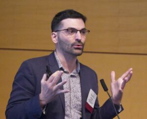
Opening the discussion, Marcel van den Brink, MD, PhD, president of City of Hope Los Angeles and City of Hope Medical Center spoke about the role of the intestinal microbiome in cancer immunotherapy.

Marcel van den Brink, MD, PhD [Nick Fetty/The New York Academy of Sciences]

He began by describing the history of graft versus host disease (GVHD), pointing out that while early work from the 1970s suggested that germ free mice had reduced instance of GVHD following transplants, more current work has pointed to the intestinal microbiome as an immune system modulator.

“Protection of the commensal anaerobes is beneficial,” he said. He explained that Enterococcus has a habit of dominating a population with reduced diversity, pointing out that it “happens very frequently within the context of allogenic transplant, and again is linked with graft versus host [disease].

“So we try to understand how that happens, why do you get that domination?” van den Brink described how damage to the internal lining of the gut by chemotherapy or XRT conditioning can lead to alloreactivity of immune cells. Damaged enterocytes are less able to produce lactase, leading to increase in lactose availability, which can help drive the growth of Enterococcus species, including E. faecillis—a primary species found in patients who develop GVHD. Concurrently, bile acids can have an immune suppressive effect.

He went on to share results of two published studies exploring the role of immune cells in this cycle and potential interventions. He summarized this work saying, “The protection of the commensal anaerobes is critical.

“That’s probably the easiest point that I can make, if you think about using the gut microbiome as a target to improve outcomes for cancer patients.”

As the gut is a complicated ecosystem, and there are many angles of research, the van den Brink lab is also now exploring other avenues of research that do not involve antibiotics in addition to their work with antibiotics and other therapies.

Probiotic engineering

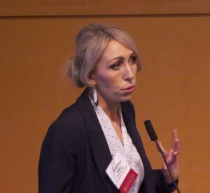
The second talk in this session, presented by Nicholas Arpaia, PhD, associate professor of microbiology and immunology at Columbia University, explored the possibility of personalized cancer immunotherapy with the use of engineered probiotics.

Nicholas Arpaia, PhD [Nick Fetty/The New York Academy of Sciences]

His work has focused on exploring the interactions between bacteria and the tumor microenvironment (TME) and how bacteria can act as a sort of Trojan horse to access the inner tumor environment.

In terms of cancer immunotherapy, Arpaia began by saying, “hopefully I’ll be able to convince you that utilizing bacteria is a potential path forward.” Bacteria, he argues, have a bright future in the field with a strong and growing research background based on the publications, companies formed, and clinical trials over the last 20 to 30 years.

He continued describing the wide scope of the field, both in how bacteria are engineered, and in how those bacteria are delivered. While the immune-oncology space has tended towards engineering payloads that modify the TME or deliver neoantigens, there are other approaches aimed at delivering toxins or modifying the metabolism within the TME. Further, Arpaia shared details on the differences between intravenous (IV), intratumor, or oral delivery. He pointed out that bacteria injected intravenously have been found in the cores of tumors. “It’s been speculated that this occurs because of the amenable conditions within the tumor.”

The question then arises, how can this behavior be beneficial to cancer therapy? “Features of bacteria themselves can activate the innate immune system,” Arpaia said. “If you then couple that with something that’s going to help activate the adaptive immune system, it gives us all the signals we need to really get long-term durable and effective responses.”

While many bacterial strategies involve the bacteria bringing specific payloads to the TME, much of his work explores a strategy of quorum-based lysis or a synchronized lysing circuit.

“Essentially what we should observe is that there’s growth, they hit a quorum threshold, so this synchronized lysis event occurs, a few of the bacteria remain, and the entire population undergoes these cyclic events again.”

Following the lysis event, what remains is “just a massive bag of innate immune stimulatory ligands.” The payload is released over and over through this synchronized lysis of the bacteria. Arpaia summed the process: “They grow, they undergo a lytic event, they grow back, and the entire process again occurs again and again.

Tumor-associated bacteria in space

The final talk seamlessly transitioned from the discussion of bacterial lysis deep within the tumor to a discussion on how the location of tumor-associated bacteria within the TME can impact therapy approaches.

Susan Bullman, PhD, associate professor of immunology at the University of Texas MD Anderson Cancer Center began her discussion by taking a step back from cancer. “What I’m going to talk about is the native colonization of tumors by bacteria, by members of our microbiome,” she said.

Susan Bullman, PhD [Nick Fetty/The New York Academy of Sciences]

She explained that her group is “particularly interested in oral gastrointestinal cancers and understanding how microbes disseminate from our microbiome and can infiltrate human tumors to modulate the TME.”

Bullman described how certain bacterial that or typically restricted to the oral cavity can migrate and infiltrate cancers throughout the gastrointestinal (GI) tract. She focused specifically on Fusobacterium nucleatum, which not only has been consistently identified in GI tract tumors, but has also been found to negatively impact patient outcomes.

“When this microbe is enriched in the tumor, patients tend to have an increased risk for relapse, metastases and overall poor prognosis,” she said. Bullman explained that there is variability between tumor types and likelihood of tumor infiltration by microbes—with GI tract tumors having a higher instance of bacterial infection. Further, there is a heterogenous distribution of the bacteria within the tumor itself and while this bacterium is not the only microbe within the tumor tissue, her work aims to understand how this species modulates the TME.

She asked, “When these microbes get into a tumor tissue or infiltrates the tumor tissue, what exactly are they doing?” She pointed out that in healthy tissue, bacteria will interact with epithelia cells and interact with the immune system, but it’s unclear what they do within the TME.

Through the use of sequencing of both tumor and bacterial cells, her lab was able to identify details on the genetic expression of tumor cells and have a better understanding of the TME. They found that just the mere presence of bacteria at all in the tumor also has a physical impact on the tumor. The bacteria have been shown to impact tumor cell density, increasing space between the human tumor cells. As a result, these cells become stressed and stay in temporary quiescence until the bacteria are removed.

“This is interesting for a range of perspectives, from an immunotherapy perspective and an immunology perspective,” she shared.

“We see that these quiescent cells, they reduce metabolism, they reduce gene expression, and they have reduced antigen presentation. So when the cancer epithelial cells are pushed into this dormant state, they become somewhat invisible to the immune system.”

From a chemotherapy perspective, this was an interesting discovery. “We know that many anti-metabolite chemotherapies that are used in the clinic, they are targeting hyperproliferative cells,” she explained.

Currently, the team is working to map the host-bacterial interactions within the TME, looking for co-localization of cells and function to better understand how the tumor responds to bacterial infection. While they are still trying to understand the mechanisms, Bullman is encouraged by the current data.

“There [are] hints towards impacts of microbes, the amount, the load of these microbes, the immune cells, the monoid cells that they’re recruiting, and their impact on immune checkpoints within the tumor microenvironment.

The post The Microbiome’s Growing Role in Cancer Immunotherapy appeared first on Inside Precision Medicine.

Alzheimer’s Targets Spotted in Circulatory System

Several genes relating to plasma proteins—particularly those involved in the immune system—have been linked for the first time with Alzheimer’s disease.

The proteome-wide association study (PWAS) strengthens the link between dementia and the immune system.

It also highlights potential therapeutic targets outside the traditional confines of the central nervous system (CNS) brain and spinal cord.

Genes relating to both the innate and adaptive immune system were linked with the neurodegenerative disease in the Science Translational Medicine study.

The present PWAS, which modeled circulating protein abundance, identified potential candidate genes, many of which are expressed broadly or predominantly outside the CNS,” reported Keenan Walker, PhD, from the National Institute on Aging in Baltimore.

“These genes exhibited a higher enrichment for adaptive immunity compared with GWAS [genome-wide association study] candidates, implying distinct mechanistic pathways operating outside the CNS.”

Data on the plasma proteome and protein quantitative trait loci (pQTLs)—genetic variants that control the expression of proteins—are increasingly becoming available.

These offer the opportunity to identify disease-related genes through their effect on protein levels. Proteins are highly relevant to diseases and the development of therapeutic targets, given their role as effectors of cellular biology.

Walker and team compared genetic and proteomic changes associated with Alzheimer’s disease by studying GWAS results from tens of thousands of cases of Alzheimer’s disease, related dementias and control individuals.

They then compared this information with genetic models of 1348 circulating proteins derived from European Americans and 1385 circulating proteins from African Americans.

Plasma cis-pQTL data identified genes associated with the neurodegenerative disease through the cis regulation of plasma protein abundance. It also highlighted several plasma proteins with potential links with dementia.

The research revealed 18 genes with potentially causal relationships with the risk of Alzheimer’s disease among people with European ancestry, six of which had not been previously identified through GWAS.

Leukocyte immunoglobulin-like receptor (LILR)B1 and signal regulatory protein alpha (SIRPA), two immunoregulatory proteins not previously implicated in GWAS, showed the strongest mechanistic link to Alzheimer’s disease in the European Americans.

“We demonstrated that a subset of the plasma proteins encoded by PWAS candidate genes may function as biomarkers, given their association with dementia risk over 8- and 20-year follow-up periods, as early as middle adulthood,” the researchers reported.

“These same plasma proteins were associated with biomarkers of [Alzheimer’s disease] pathology and a broad set of cerebrospinal fluid (CSF) proteins enriched for immune and metabolic processes relevant to [Alzheimer’s disease].

“The present PWAS, which modeled circulating protein abundance, identified potential candidate genes, many of which are expressed broadly or predominantly outside the CNS.

“These genes exhibited a higher enrichment for adaptive immunity compared with GWAS candidates, implying distinct mechanistic pathways operating outside the CNS.”

The post Alzheimer’s Targets Spotted in Circulatory System appeared first on Inside Precision Medicine.