Cellares and Sonoma Biotherapeutics Agree to Automate Engineered Treg Cell Therapy Manufacturing

Officials at Cellares and Sonoma Biotherapeutics say their companies will automate the manufacturing of SBT-77-7101, an engineered regulatory T cell (Treg) therapy for autoimmune and inflammatory diseases. The product is in Phase I clinical development for poly-refractory rheumatoid arthritis (RA) in patients who have exhausted all available treatment options.

Cellares plans to translate SonomaBio’s SBT-77-7101 manufacturing process onto its Cell Shuttle and automate in-process and release testing through the Cell Q™ quality control system. The company operates its first commercial-scale Smart Factory in Bridgewater, NJ, with additional facilities under construction in Europe and Japan.

“Tregs are uniquely sensitive to the manufacturing process. Cellares brings the Cell Shuttle platform, and global infrastructure to help us deliver on our clinical ambitions at scale for the hardest-to-treat RA patients.” said Stephen Dilly, PhD, president, CEO, and board chair of Sonoma Biotherapeutics.

“Every new cell therapy modality we bring to the Cell Shuttle and Cell Q expands what is possible for the field and for patients in need,” added Fabian Gerlinghaus, co-founder and CEO of Cellares. “Tregs are among the most technically demanding cell types to manufacture reliably. We are honored to partner with SonomaBio and demonstrate that our platform can directly translate to Tregs.

“SonomaBio has developed one of the most advanced Treg programs in the clinic, and we look forward to contributing to their clinical success as they bring this groundbreaking therapy to patients.”


The post Cellares and Sonoma Biotherapeutics Agree to Automate Engineered Treg Cell Therapy Manufacturing appeared first on GEN – Genetic Engineering and Biotechnology News.

Genome-Scale CRISPRi Atlas Maps Gene Function Across Human iPSCs

A new genome-scale atlas is offering an unprecedented look at how individual genes shape the transcriptional landscape of human induced pluripotent stem cells (iPSCs). Published in Nature Biotechnology, the resource catalogs the effects of perturbing 11,692 expressed genes across more than 2.5 million single cells, creating a reference framework for understanding how pluripotent identity is maintained and regulated.

The study is titled, “A genome-scale CRISPRi perturbation atlas of human induced pluripotent stem cells.”

Human iPSCs can differentiate into virtually any cell type, yet the functions of most genes within this state remain poorly understood. Prashant Mali, PhD, senior author and professor of bioengineering at UC San Diego, said the team set out to fill that gap by systematically switching off genes one by one using CRISPR interference (CRISPRi) and measuring the resulting transcriptome-wide changes. “The result is a kind of reference atlas; it’s a way to look up what perturbing almost any gene does to a stem cell’s behavior, measured here as the impact on its whole transcriptome,” Mali said.

The dataset captures how gene perturbations cluster into shared molecular signatures, revealing functional relationships among protein complexes, metabolic pathways, and self-renewal genes. By correlating transcriptional phenotypes across thousands of perturbations, the researchers reconstructed a map of the pluripotent state that recapitulates known regulatory modules while surfacing previously unrecognized ones.

Exploring the atlas led the team to identify new regulators of stem cell biology. They uncovered ZBTB41 as a metabolic factor and RNF7 as a contributor to pluripotency regulation, validating both through metabolic tracing, immunofluorescence, and protein–protein interaction assays. The resource also enabled a genome-scale screen of A‑to‑I RNA editing modulators, revealing DBR1 as a potent regulator of adenosine-to-inosine conversion.

Co-first author Yesh Doctor, a bioengineering PhD student in Mali’s lab, described the atlas as a “hypothesis engine” for stem cell researchers. Instead of running thousands of perturbation experiments, scientists can now query the open-access map to identify candidate genes involved in differentiation, metabolism, or disease-relevant pathways. “Scientists can use it to look up the functions of genes and build hypotheses on them instead of having to run the experiments themselves,” Doctor said.

Beyond basic biology, the team sees the atlas as a foundation for computational modeling. The scale and consistency of the dataset make it well suited for training AI systems aimed at predicting genotype–phenotype relationships. “These comprehensive, genome-scale screens enable generation of reference maps that are not just invaluable for basic science discovery, but also an important resource for powering future computational and AI tools for genotype-phenotype prediction,” Mali said.

The open-access atlas is available here, providing a new reference point for understanding how genes shape human stem cell identity and offering a tool for virtual disease modeling and target discovery.

The post Genome-Scale CRISPRi Atlas Maps Gene Function Across Human iPSCs appeared first on GEN – Genetic Engineering and Biotechnology News.

Analytical Challenges for Antibody-Drug Conjugate (ADC) Manufacturing

Antibody-drug conjugates (ADCs) offer a compelling promise: delivering a cytotoxic payload directly to a tumor cell while sparing the rest of the body from harm. Composed of a tumor-targeting antibody, a cytotoxic agent, and a chemical linker, ADCs combine the selectivity of monoclonal antibodies that bind to tumor-specific antigens with the cytotoxic potency of small-molecule drugs. These therapeutics have the potential to target tumors while reducing systemic toxicity, opening new treatment pathways for many types of cancer. Recent clinical data, including encouraging Phase I findings in platinum-resistant ovarian cancer, validate that potential and fuel the field’s remarkable growth.

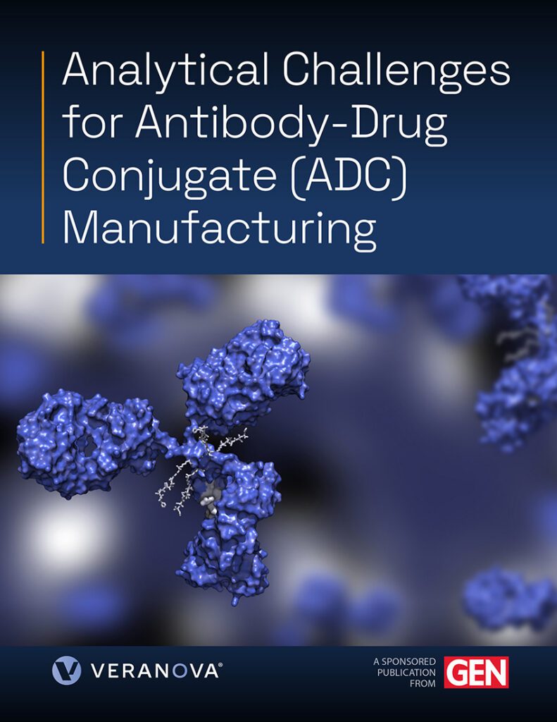
Analytical Challenges for Antibody-Drug Conjugate (ADC) Manufacturing cover

Despite the promise of ADCs, their unique structure complicates manufacturing, characterization, and regulatory assessment. Developers must carefully consider the interplay between the antibody, linker, and payload to optimize therapeutic efficacy and safety. The drug-to-antibody ratio (DAR), conjugation site specificity, impurity profile, and linker stability all influence an ADC’s pharmacokinetics, pharmacodynamics, and ultimately its clinical safety profile. Robust analytical methods are therefore critical throughout the development process to ensure both safety and efficacy.

Given the hybrid nature of ADCs, regulatory expectations are still evolving. To navigate this uncertainty, analytical risk management is essential. Moreover, cross-functional collaboration among analytical scientists, process development teams, regulatory experts, and quality assurance professionals is key to ensure that early-stage methods are sufficiently robust and scalable for commercial manufacturing.

Success in this environment requires deep analytical expertise, robust quality-by-design frameworks, and development partners who understand the full arc from early-stage linker-payload synthesis through GMP-compliant manufacture. It requires the capability to handle highly potent compounds safely, to purify structurally complex intermediates at scale, and to translate rigorous quality control into processes that are commercially viable.

This collection of articles and expert perspectives explores the critical challenges shaping ADC development today, from analytical strategy and impurity control to linker technology innovations and evolving regulatory standards. It also examines the collaborative expertise needed to bring these transformative therapies to patients.

The post Analytical Challenges for Antibody-Drug Conjugate (ADC) Manufacturing appeared first on GEN – Genetic Engineering and Biotechnology News.

New Lung Model Reveals RSV Immune Response, Points to Better Treatments

Researchers at University College London (UCL) Great Ormond Street Hospital for Children (GOSH) have built a new lab model of infant lungs to show why respiratory syncytial virus (RSV) makes infants so much sicker than adults, and to allow them to test new treatments. The miniature model of a baby’s airways was created using pediatric airway cells, blood vessel cells, and neutrophils (a type of white blood cell that acts as the immune system’s primary response to infection).

Studies using the new model suggest that future therapies for RSV should target both the virus and its immune response to ensure babies get the best possible outcomes. Research lead Claire Smith, PhD, at UCL Great Ormond Street Institute of Child Health, said, “This model allows us to watch early immune responses unfold and study them in a human setting that reflects the infant airway. That’s something animal models often struggle to capture, especially when it comes to age-specific effects.”

Senior and corresponding author Smith and colleagues reported on their findings in Nature Communications in a paper titled “Neutrophil myeloperoxidase as a functional biomarker for RSV severity: implications for in vitro therapeutic screening.” In their report they concluded, “These findings identify neutrophil–epithelial interactions as a useful target for intervention and support the use of physiologically relevant human models to accelerate the development of therapies that limit immunopathology while preserving antiviral defense.”

RSV is the biggest cause of severe respiratory tract infections in infants and young children, resulting in over three million hospital admissions worldwide every year. RSV infection causes wheezing and breathing difficulties, and in the worst cases infants end up in intensive care. Despite this, treatment options for severe RSV disease remain extremely limited. “The lack of accessible, effective therapies highlights the need for continued advancements in RSV treatment and prevention,” the authors wrote.

During RSV infection neutrophils are rapidly mobilized to the lungs and play a key role in virus-targeting host defenses, the investigators continued. However, excessive neutrophil infiltration and activation can also contribute to airway inflammation, epithelial damage, and disease severity. “Understanding neutrophil behavior and activation during RSV infection, including during their migration across the airway epithelial barrier, is crucial for developing therapeutic strategies to mitigate pathological inflammation without compromising the antiviral response,” they suggested.

For their reported study the team aimed to create an in vitro model that recapitulates key clinical outcomes of infants with RSV bronchiolitis. To do this they established an air-liquid interface (ALI) system that incorporated pediatric airway epithelial cells, endothelial cells, and neutrophils from adults, to mirror the blood-airway barrier. “Differentiated airway epithelial cells (AECs) cultured at the air–liquid interface (ALI) provide a physiologically relevant platform to study neutrophil migration and the effect of antiviral treatments on this process,” they note. To compare with an adult response to RSV, the research team also made a model of an adult’s airways.

When the models were infected with RSV, the team found that the pediatric airway cells attracted far more white blood cells than did the adult airway cells. This influx can block an infant’s small airways and make it harder for them to breathe.

Neutrophils normally circulate in the blood but enter lung tissue in response to infection. In the baby airway model, researchers found that the neutrophils that entered the lung tissue were more activated and triggered a stronger inflammatory reaction than in the adult model. This effect was dependent on the immune cells physically moving through the infected tissue, not just responding to chemical signals released by it, making this type of model essential for study.

The results suggest that it is the infant airway itself, not just the virus, that ramps up the immune response and causes damage to the lungs. First author Machaela Palor, PhD, at UCL Great Ormond Street Institute of Child Health, said: “These findings help explain why RSV is often much more severe in infants than in adults. The pediatric airway actively shapes how immune cells behave during the infection.”

The researchers tested two antiviral drugs (remdesivir and RSV604). they found that both stopped the virus from multiplying, but only RSV604 also calmed the overactive immune response, reducing levels of a key inflammatory protein myeloperoxidase (MPO) released by white blood cells—high levels of which are linked to more severe RSV disease in babies. Remdesivir had no effect on this, suggesting that not all antivirals are equal when it comes to protecting the infant airway from immune-driven damage. “While both drugs reduced viral load, only RSV604 attenuated MPO expression,” the team stated.

The findings suggest that treating severe RSV in babies may require more than just stopping the virus—it may also be important to calm an overactive immune response. “This model suggests that MPO could be useful as a readout of therapeutic efficacy,” the team stated. “Targeting neutrophil-driven inflammatory pathways may be critical for reducing pathology in infant RSV infection.”

The researchers hope their findings and the new approach to research on RSV will accelerate the development of treatments better tailored to infants. “Our model gives us a way to assess both sides of the problem at once,” Smith said. “We can not only ask whether the drug stops the virus but also whether it helps control immune response in the infant airway. “This work reinforces the idea that age matters in respiratory infection. Understanding how infant airways shape immune responses will be key to designing safer and more effective RSV treatments.”

The post New Lung Model Reveals RSV Immune Response, Points to Better Treatments appeared first on GEN – Genetic Engineering and Biotechnology News.


Why Navigantis thinks it can win the stroke telesurgery race

Navigantis says it has what it takes to win the race to treat stroke patients remotely with its interventional robotic platform VASCO, which is already being used in a clinical trial for neurointerventional procedures. Stroke telesurgery is one of the biggest opportunities in medtech. Major OEMs and startups barely out of stealth mode are racing…

The post Why Navigantis thinks it can win the stroke telesurgery race appeared first on Medical Design and Outsourcing.

FDA releases draft MDUFA VI agreement outlining proposed legislation and program changes

A faster pre-submission track, earlier communication for de novo applicants and an “international harmonization” pilot program are among proposed improvements and changes featured in the FDA’s latest draft Medical Device User Fee Amendments (MDUFA) agreement. The agency recently released a draft commitment letter, detailing its proposed agreement with the medtech industry for MDUFA reauthorization. If…

The post FDA releases draft MDUFA VI agreement outlining proposed legislation and program changes appeared first on Medical Design and Outsourcing.

Auxilium bioprints kidney and liver tissues in space for the first time

Auxilium Biotechnologies says it bioprinted kidney and liver tissues aboard the International Space Station in the “first demonstration of a scalable, multi-product biomanufacturing platform in space.” The San Diego-based biotech and device developer used its Auxilium Microfabrication Platform (AMP-1) 3D bioprinter for the first-of-its-kind mission, which also included orbital bioprinting of cartilage tissue and nerve…

The post Auxilium bioprints kidney and liver tissues in space for the first time appeared first on Medical Design and Outsourcing.

Immune Evasion Uncovered by Virome-Wide Ubiquitin Ligase Discovery

In a new study published in Science titled, “Virome-wide ubiquitin ligase discovery reveals diverse mechanisms of immune evasion,” researchers from Harvard Medical School (HMS) have uncovered how viruses hijack cells’ garbage-disposal systems to evade immune attack. 

The study applies ORFeome, a tool that broadens the scale by which researchers study viral proteins. The advance proposes to speed basic discoveries in virology, inform the development of new vaccines and treatments to protect against emerging pathogens. 

“This library reveals how viruses manipulate human cells on a scale that simply wasn’t possible before,” said Stephen Elledge, PhD, professor of genetics and medicine at Harvard Medical School and senior author of the study. “We believe it changes virology from studying one virus at a time to discovering the common strategies and surprising innovations that viruses have evolved, providing a powerful new foundation for understanding emerging viral threats.” 

ORFeome and other ORF libraries are named after open reading frames, DNA sequences that encode proteins. Previous viral ORF libraries from other groups focused on individual viruses or virus families that contained 100 or 200 sequences each. The new ORFeome contains about 13,000 physical DNA sequences, or constructs, that code for about 9,000 proteins from 513 different viruses, including Andes hantavirus, Ebola virus, and Zika virus. 

“Most viruses have never been studied in detail, yet evolution has already performed countless experiments for us. This library gives us a way to read the results of those experiments across the viral world,” said Elledge, who is also a Howard Hughes Medical Institute (HHMI) Investigator. 

The team attached a genetic barcode to each ORF, allowing researchers to conduct studies of all 13,000 ORFs at once. 

“We can insert the sequences into a population of cells, ask questions like which ones cause the cells to grow better or less, and then identify those by their barcodes when the experiment is finished,” said Colin O’Leary, PhD, HMS research fellow and co-author on the study. “It hasn’t been possible before to do genetic screens like this with viral proteins.” 

The team will make the ORFeome freely available for the research community. Elledge and colleagues implemented a flexible design to enable application to other model systems and experiments. 

The team conducted genetic screens in three cell types, searching for viral proteins that affect cell proliferation, stop cells from presenting antigens to trigger the immune system to attack, or block the effects of interferon. Results uncovered more than 700 viral proteins that contribute to at least one of those actions. 

The study opens opportunities to design drugs that hinder viral activity while sparing normal function. 

The post Immune Evasion Uncovered by Virome-Wide Ubiquitin Ligase Discovery appeared first on GEN – Genetic Engineering and Biotechnology News.

STAT+: All eyes on data on Biogen’s tau therapy

Want to stay on top of the science and politics driving biotech today? Sign up to get our biotech newsletter in your inbox.

Lots happening in London these days, my colleague Andrew Joseph reports: At a pancreatic cancer meeting, researchers were buzzing about the implications of daraxonrasib, and at a major Alzheimer’s meeting, tau therapies are taking center stage.

Back here in the U.S., HHS has begun to craft national guidance on safely tapering SSRIs, and the FDA just cleared Sanofi’s wearable injector for a myeloma drug.

Continue to STAT+ to read the full story…