A new startup has found that the Chinese biotech industry is good for more than obesity drugs and cancer therapies.
The company, Tortugas Neuroscience, launched Tuesday with plans to develop two schizophrenia and tinnitus drugs licensed from Chinese drugmaker Jiangsu Hansoh Pharmaceutical Group. The startup will also test two other medicines for focal epilepsy and encephalopathies that were originally created by Japanese pharmaceutical company Eisai Co. Ltd.
Tortugas has $106 million from Cure Ventures, The Column Group, and AN Ventures to begin testing the drugs in mid-stage trials in the U.S.
Freudenberg Medical, a Kaiserslautern, Germany-based contract design manufacturing partner, launched CleanAssure, a new ISO Class 5 controlled cleanroom designed to deliver clean and sterile single-use assemblies for biopharmaceutical customers, according to the company.
Freudenberg Medical manufactures silicone and thermoplastic elastomer (TPE) tubing for bioprocessing and critical fluid transfer. The company specializes in seamless, overmolded single-use assemblies used in vaccine production, cell cultivation, fluid transfer, and fill-finish operations.
The controlled cleanroom enables customers to receive ready-to-use washed, dried, and gamma sterilized single-use assemblies with the highest product quality, sterility, and process consistency, noted Rudi Gall, vp, global pharma, Freudenberg Medical.
“CleanAssure allows us to support our customers beyond component manufacturing,” he said. “By integrating controlled cleaning and sterilization into our single-use assembly services, we help reduce contamination risk, streamline validation activities, and support a reliable supply for our customers. We can now support customers with their entire component value chain and allow them to focus on their core manufacturing capability.”
Freudenberg’s cleaning process uses ultrapure water and air, operating within ISO 5 conditions. The water is produced using a multi-stage filtration process, resulting in high-purity water specifically suitable for pharmaceutical applications.
Key biopharma industry challenges
Biopharmaceutical manufacturers increasingly rely on single-use systems but face ongoing challenges related to cleaning validation, contamination risk, and production downtime. Customer-managed cleaning processes are often time-intensive, costly, and require additional resources while directly impacting supply reliability, according to Gall.
The company explained that its controlled cleaning environment addresses these challenges by reducing cross-contamination risk through tightly controlled ISO Class 5 processing; alleviating customer cleaning validation burden by delivering assemblies washed and sterilized under cGMP, validated conditions; minimizing production downtime by removing cleaning as a process step; and supporting a consistent, reliable supply of high-quality single-use assemblies.
Freudenberg will be attending INTERPHEX New York, April 21–23, Booth 1673, to exhibit its new products and services.
How does HIV, armed with only nine genes, manage to hijack the immune system so effectively? For decades, researchers have known that the virus depends on human proteins to enter, replicate, and persist—yet the full roster of those host factors has remained elusive. One major reason: most HIV studies have relied on immortalized cell lines rather than the primary CD4+ T cells the virus actually infects in the body. As a result, scientists have lacked a comprehensive picture of how real human T cells respond when HIV attacks.
A new study from Gladstone Institutes and the University of California, San Francisco (UCSF), changes that. In the study, titled “Systematic Discovery of Pro- and Anti-HIV Host Factors in Primary Human CD4+ T Cells” and published in Cell, researchers report the first genome‑wide map of human genes that either promote or restrict HIV infection in primary human CD4+ T cells, offering a long‑sought blueprint of the host–virus interface.
“HIV has been a global crisis for over 40 years,” said Alex Marson, MD, PhD, director of the Gladstone‑UCSF Institute of Genomic Immunology and senior author of the study. “By studying human T cells, which are the primary target of the virus, we’ve finally mapped the genes—many of which were previously unknown—that influence whether or not they can be infected by HIV.”
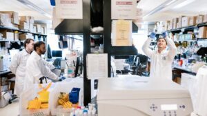
Scientists in the Marson Lab at Gladstone Institutes have opened a new door to understanding HIV by creating the first genetic roadmap of how the virus interacts with real human cells. [Gladstone Institutes]
Achieving this required overcoming a fundamental technical barrier. “One challenge of using real human T cells for research is they’re very difficult to infect with HIV; out of a whole dish of cells, typically only one or two percent would get infected,” said first author Ujjwal Rathore, PhD. After years of optimization, the team pushed infection rates to roughly 70%, enabling genome‑scale CRISPR perturbations in primary cells for the first time.
With that platform in hand, the researchers performed orthogonal genome‑wide CRISPR activation (CRISPRa) and CRISPR knockout (CRISPRn) screens in CD4+ T cells, systematically testing nearly every human gene. Disrupting genes revealed those HIV depends on, while overactivating genes exposed natural antiviral defenses that HIV normally suppresses. “Over‑activating the genes gave us a wealth of information,” said co–first author Eli Dugan, a PhD candidate in Marson’s lab. “We discovered natural antiviral proteins that were previously invisible because the virus could effectively silence them.”
Across both screens, the team identified hundreds of host factors that shape HIV infection. Among the most striking were two previously unrecognized antiviral proteins: PI16 and PPID (Cyp40). “PI16 interacts with host factors involved in HIV fusion and inhibits viral entry, whereas PPID, a paralog of the proviral cyclophilin CypA, binds capsid and reduces nuclear import of the HIV core,” wrote the authors. Targeted mutagenesis, along with structural modeling and evolutionary analyses, pinpointed residues essential for PPID’s restriction activity, and engineered variants were up to tenfold more potent, according to Dugan.
To test whether these defenses could counter real‑world viral strains, the team collaborated with HIV pioneer Jay Levy, MD, who provided isolates from the early AIDS epidemic. Elevated levels of PI16 or PPID restricted even these aggressive HIV strains.
“This was the first genome‑wide effort to show how human genes affect HIV infection in cells taken directly from human blood samples,” said Nevan Krogan, PhD, director of the HIV Accessory and Regulatory Complexes (HARC) Center. “Our findings could eventually lead to new treatments that help the body’s immune system resist the virus.”
Beyond identifying antiviral factors, the study offers the potential for a powerful new platform for probing HIV latency—the persistent reservoir that evades antiretroviral therapy. “Now, we have the platform to ask the biggest questions in the field,” Rathore said, “and hopefully learn how to eliminate hidden HIV that current drugs can’t reach.”
Damage to the liver in patients developing end-stage liver disease has become too severe for the organ’s normally extraordinary regenerative capacity to repair or compensate for that damage. Once this point of no return has been reached the only option is an organ transplant. However, donor livers are in high demand and very limited supply.
Ambitious efforts are on the way that eventually could enable the engineering of entire implantable liver organs. However, the maximum size of laboratory-engineered liver constructs remains limited and cannot yet provide therapeutic benefits for patients. A research team at the Wyss Institute at Harvard University, Boston University, and MIT has now approached this important problem from a different angle.
“We asked if it would be possible to first implant a small-scale liver construct and then drive it to expand in the body following its engraftment,” said Christopher Chen, MD, PhD, a Wyss Institute core faculty member and the William Fairfield Warren Distinguished professor of biomedical engineering and director of the Biological Design Center at Boston University. “A sufficiently grown, functional ‘satellite liver’ could immediately relieve the metabolic burden in a damaged liver and help bridge the time until a transplant becomes available.”
Chen co-led the research together with associate faculty member Sangeeta Bhatia, MD, PhD, who is the John J. and Dorothy Wilson Professor of Health Sciences and Technology and of Electrical Engineering and Computer Science at the Koch Institute for Integrative Cancer Research at MIT, and a Howard Hughes Medical Institute investigator. Chen is also a leader of the Wyss Institute’s 3D Organ Engineering Initiative, and team lead of the recently awarded ARPA-H PRINT-supported ImPLANT project, which focuses on whole organ liver engineering at the Wyss and collaborating institutions.
The project, spearheaded by Amy Stoddard, PhD, (MIT ’25), who developed the approach in her doctoral research and then as a postdoctoral fellow, integrates tissue engineering and synthetic biology tools in a genetic strategy the team has named “bioengineered on-demand outgrowth via synthetic biology triggering,” or BOOST. By specifically rewiring the gene expression of primary liver hepatocytes and supportive fibroblast cells, the scientists were able to effectively switch on a tissue growth program in small, engineered liver constructs after their implantation into mice.
“Using engineered liver tissue as a proof-of-concept application, we integrated synthetic biology and tissue engineering tools to build liver tissues that can be expanded on-demand after implantation in vivo,” the team reported in their published paper in Science Advances, which is titled “Synthetic control of implanted engineered liver tissue growth.” In the paper they concluded “In this study, we define the first steps toward an unconventional approach to cell therapy scale-up: engineering a small construct and then inducing it to grow in situ … “This strategy, which we have named BOOST, could provide several key advantages, including circumventing the need for large quantities of cellular raw materials and bypassing the formidable challenge of generating a rapidly perfusable construct that can survive the engraftment period.”
The authors wrote, “Organ transplant is currently the only curative treatment for patients with end-stage organ failure, yet this therapy is inaccessible to many due to the paucity of organs available for transplant.” And while significant progress has been made in the field of engineering tissue-based cell therapies that could represent alternatives, or bridges to transplant, they acknowledge, “… scaling of these constructs to sizes of therapeutic relevance remains a barrier to clinical translation.”
In order to address current challenges associated with fabrication, Chen and colleagues looked at the problem from different angle, asking whether it would be possible to first implant a small-scale construct and then trigger it to expand in situ, after its engraftment into the host.
To be able to induce growth of an implanted small liver constructs in situ within a recipient’s body the researchers first needed to identify the relevant cues that would allow them to do so. “A key first step toward this method of in situ scale-up would be the successful control of cellular growth within the engineered construct after engraftment,” they wrote. Since liver growth is known to be regulated by soluble growth factors (GFs), Stoddard screened a collection of candidate factors to identify those that, when added to cultured human primary hepatocyte cells (HEPs), had the strongest growth-inducing effects.
The genetic “BOOST” strategy integrates tissue engineering and synthetic biology tools to enable on-demand liver growth inside the body. By specifically rewiring the gene expression of primary liver hepatocytes and supportive fibroblast cells, a tissue growth program is switched on in a small, engineered liver construct after its implantation into recipients and upon addition of an inducing agent (shown as a pill). As a result, the hepatocytes in the construct start and continue to proliferate until a desired construct size has been reached and the inducing signal is not provided anymore. In mice, BOOST resulted in robust and healthy liver growth. [Wyss Institute at Harvard University]
“We ended up with a set of four growth factors, HGF, TGFa, WNT2 and RSPO3, that potently induced sparsely scattered HEPs to grow in the culture dish,” said Stoddard. “But when we tested whether they could do the same in 3D liver tissues consisting of densely packed HEPs and fibroblasts, they turned out to be ineffective. This led us to hypothesize that there must be an additional mechanism at work in human HEPs that inhibits cell proliferation in high-density conditions.”
The team homed in on a protein, YAP, that senses mechanical signals, and which was known to move from cells’ cytosol to their nucleus in low-density conditions to help express genes involved in cell proliferation. However, in high-density conditions when cells are compressed, YAP is degraded in the cytosol, which prevents the activation of those target genes and restricts proliferation.
“Importantly, when we overexpressed a non-degradable version of YAP in HEPs, which also reaches the nucleus in high-density conditions to partake in gene regulation, we successfully overrode this density checkpoint in HEPs,” Stoddard said. “Interestingly, we found that HEPs needed to be stimulated with both YAP and GFs in order to grow in densely packed 3D liver tissues.”
Toward the goal of safely inducing and controlling HEP proliferation in a living organism, and eventually human patients, the researchers deployed synthetic biology tools to locally install control of these signaling pathways in HEPs and fibroblast cells within the engineered 3D liver tissues themselves. “We set out to engineer a synthetic biology toolkit capable of locally modulating growth factor and YAP signaling within engineered liver tissue, enabling on-demand control of proliferation even after implantation,” they noted.
The team engineered fibroblast cell lines that each secreted one of the four GFs, and HEPs that expressed the non-degradable YAP protein. And they made the expression of all proteins doxycycline (DOX)-inducible. They determined in time course experiments that a continuous seven-day treatment with DOX led 3D liver tissue composed of engineered cells to robustly expand in size and cell numbers in the culture dish. On DOX removal the HEPs reverted back to a non-proliferating state.
However, Stoddard noted, “… when we compared the gene expression of single cells in BOOST-engineered, DOX-induced 3D liver tissue to that of cells in non-engineered or BOOST-engineered, non-induced 3D liver tissue, we noticed that the expansion came with a trade-off: high proliferation rates went hand in hand with a less functional HEP state. While we believe this is a natural trade-off seen in a wide variety of biological settings, we hope to be able to address this in the future, recognizing that the liver also has native re-functionalization signals to harness.”
The litmus test for BOOST-engineered growth in 3D liver tissues was to see whether they would similarly expand following their implantation into living mice that were systemically treated with DOX for the same seven-day duration. Experiments showed that the implanted tissue exhibited a striking 500% increase in proliferation with a doubling of the engineered HEPs alone, and was vascularized to accommodate the metabolic demands of the expanded tissue. The tissue implants were also well tolerated by the mice, with no signs of fibrosis due to invading immune cells and fibroblast inflammation, or of tumor growth.
“These results were particularly exciting to us,” said Stoddard. “Prior to our work, injury to the host liver has always been required to trigger hepatocyte engraftment and proliferation. Here we were able to relieve this dependence, and trigger on-demand growth of implanted liver tissue in a completely healthy host.”
In the future, the team will explore the capacity of BOOSTed liver tissue to rescue the host in the setting of liver injury. “Our BOOST strategy lays the foundation for a future when solid organ cell therapies can be controlled non-surgically according to the needs of patients and their physicians,” Bhatia noted. “Beyond treating liver disease, the premise of BOOST could be applied to other engineered tissue therapeutics that are similarly constrained by challenges associated with tissue scale-up, such as engineered heart or pancreatic tissue to address serious diseases.”
In their paper the authors concluded, “… this work serves as an exciting proof-of-concept demonstration that scale-up of tissues via growth could be possible … Together, this work helps lay the foundations for a future of ‘smart’ tissue therapeutics that can be scaled to a patient’s needs and thereby offer treatment for numerous, previously incurable, diseases.”
Eli Lilly has agreed to acquire Kelonia Therapeutics for up to $7 billion, the companies said today, in a deal that would bolster the buyer’s oncology pipeline with an early clinical phase lentiviral in vivo chimeric antigen receptor T-cell (CAR T) therapy under study in relapsed/refractory multiple myeloma.
Kelonia’s lead program KLN-1010 is a one-time intravenous gene therapy designed to generate anti-B-cell maturation antigen (BCMA) CAR T cells, targeting the BCMA protein expressed on the surface of multiple myeloma cells.
In December at the American Society of Hematology (ASH) 2025 Annual Meeting, Kelonia presented positive early clinical data for KLN-1010 from the Phase I inMMyCARTM trial (NCT07075185). The data showed the CAR T therapy to have 100% minimal residual disease (MRD)-negative response rate across four patients, all of whom remained in response through the longest follow up of five months.
Those and other results, according to the company, provided initial clinical validation of KLN-1010 and demonstrated promising tolerability. In January, Kelonia won FDA clearance for an investigational new drug (IND) application for KLN-1010, enabling the trial to expand from Australia into multiple clinical sites across the United States.
“The early clinical data for KLN-1010 are highly encouraging, both as a potential step forward for patients with multiple myeloma and as proof of concept for Kelonia’s platform,” Jacob Van Naarden, executive vice president and president of Lilly Oncology and head of corporate business development, said in a statement.
Investors appeared more sanguine about the Kelonia acquisition as Lilly shares were all but flat in early Monday trading as of 11 a.m. ET, to $927.16 from Friday’s close of $927.03. Kelonia is privately held.
KLN-1010 applies the company’s in vivo gene placement system (iGPS®), which uses engineered lentiviral-based particles designed to efficiently and selectively enter T-cells inside the body, enabling a patient’s own body to generate CAR T therapies designed to treat underlying disease.
Lilly and Kelonia reason that KLN-1010 could transform treatment of multiple myeloma by eliminating challenges associated with both ex vivo patient-specific cell therapy manufacturing, and pre-administration chemotherapy.
“Autologous CAR T therapies have meaningfully improved outcomes for patients with various cancers, but significant manufacturing, safety, and access barriers mean that only a fraction of eligible patients actually receive them,” Van Naarden added. “Kelonia’s in vivo platform has the potential to change that by delivering rapid, durable responses in a far simpler, off-the-shelf format.”
Kelonia marks Eli Lilly’s fourth announced acquisition of a smaller biotech this year:
And in January, Lilly inked a $1.2 billion acquisition of Ventyx Biosciences, an NLRP3-targeting oral drug developer focused on inflammatory diseases.
Behind the deals
Behind all the deals is the pharma giant’s desire to capitalize on the billions of dollars it is generating from sales of its obesity and diabetes drugs based on glucagon-like peptide 1 (GLP-1) receptor analysts alone or in tandem with a glucose-dependent insulinotropic polypeptide (GIP). Lilly markets tirzepatide, a GLP-1/GIP dual agonist, in obesity as Zepbound® ($13.542 billion in 2025 sales) and in diabetes as Mounjaro® ($22.965 billion).
Lilly stands to generate even more in obesity-related sales in coming years once it brings to market its oral obesity drug Foundayo (orforglipron), a small molecule GLP-1 receptor agonist—though analysts predict the drug’s 2026 sales will likely be lower than once expected because of the price war Foundayo faces competing head to head with Lilly’s arch-rival in obesity drugs, Novo Nordisk. In December, Novo Nordisk got a jump on Lilly when the Danish biotech giant won FDA approval for oral Wegovy® (semaglutide), a once-daily 25 mg GLP-1 receptor agonist tablet indicated for chronic weight management.
A Lilly buyout of Kelonia could compel Johnson & Johnson to take a closer look at acquiring Legend Biotech, Kostas Biliouris, PhD, a managing director on the biotechnology research team of Oppenheimer, wrote Sunday in a research note. He cited the fact J&J’s Janssen Biotech successfully partnered with Legend to develop Carvykti® (ciltacabtagene autoleucel), a B-cell maturation antigen (BCMA)-directed CAR T-cell therapy indicated for adults with relapsed or refractory multiple myeloma who have received at least one prior line of therapy. Carvykti generated $1.877 billion in sales last year, up nearly double (96%) from $963 million in 2024.
Also, Biliouris cited the presence in Legend’s pipeline of LUCAR-G39D, a clinical in vivo CAR T program designed to treat B-cell non-Hodgkin’s lymphoma by targeting CD19xCD20. LUCAR-G39D showed positive first-in-human safety and efficacy data from a Phase I trial (NCT06395870) at ASH last December.
“We believe in vivo CAR T technology has strong potential, as treatment process is fast and circumvents the need for lymphodepletion, but think it will likely take ~6-8years before safety/durability questions are addressed, and regulatory approval is granted,” Biliouris predicted.
Lilly has agreed to acquire Kelonia for $3.25 billion upfront plus up to $3.75 billion in future payments tied to achieving specified clinical, regulatory, and commercial milestones. The acquisition deal is subject to regulatory approvals and other customary closing conditions, and is expected to close in the second half of 2026.
Upon closing, Lilly said, it will determine how to account for the transaction in accordance with Generally Accepted Accounting Principles (GAAP), then reflect the deal in future financial results and financial guidance.
“Kelonia’s leadership in advancing the immense promise of in vivo cell therapy is unmatched, extending its reach and impact beyond the traditional boundaries of personalized medicine,” Kelonia CEO Kevin Friedman, PhD, stated. “We have demonstrated the ability to achieve deep multiple myeloma remissions with significantly reduced complexity and cost relative to ex vivo CAR T-cell approaches.”
“In combination with Lilly’s strengths, our in vivo iGPS platform is positioned to broaden the reach of cell therapy beyond the current CAR T landscape in hematologic malignancies and to transform treatment across a far wider range of cancers and other serious diseases,” Friedman added.
SAN DIEGO – Researchers from Kindai University in Japan have developed a machine learning model that accurately predicts the origin of diverse cancer types in patients with cancers of unknown primary (CUP) by analyzing CpG-based DNA methylation. Results showed that the model correctly identified the cancer type in about 95% of cases in the test cohort, and achieved 87% accuracy when applied to an independent validation cohort from 31 cases representing 17 different cancer types. The work was presented at the American Association for Cancer Research (AACR) Annual Meeting.
“Our findings suggest that DNA-based approaches can help identify where a cancer may have started, even when the original tumor is not visible,” said Marco A. De Velasco, PhD, a faculty member in the department of genome biology at Kindai University in Japan.
CUP are metastatic malignancies in which the primary cancer site could not be identified. These cancers are often associated with poorer outcomes, as patients are typically treated with broad, nonspecific chemotherapy regimens rather than therapies targeted to a specific cancer type.
Approximately only 15-20% of patients with CUP show features that allow site-specific therapies. Patients receiving site-directed therapy can survive up to 24 months, compared with six to nine months for those receiving standard treatment.
Patterns in tumor biology, such as gene activity or chemical modifications to DNA, can differ between cancer types and persist even after the cancer has spread and guide development of these therapies. While some methods have shown promise, they have yet to demonstrate clear survival benefits in clinical trials.
The model was developed using methylation data from nearly 7,500 patients with 21 different cancer types obtained from The Cancer Genome Atlas Program and other public datasets. Using machine learning, the researchers identified CpG methylation and built methylation profiles that were associated with different tumor types.
Del Velasco emphasized that the study achieved high accuracy in predicting the origin of diverse cancer types using a small subset of DNA markers, about 1,000 CpG regions selected from hundreds of thousands across the genome. “This is important because it shows that we can simplify complex molecular data while still maintaining strong predictive performance,” he said.
As a limitation, the model was developed using cancers with known origins, rather than true CUP. Testing in CUP patients is important to understand how well the model performs in clinical settings. Additionally, not all tumors are easily accessible for genetic testing, particularly tumors in advanced stage. Looking ahead, the authors aim to adapt and evaluate the model using blood-based biopsy to analyze circulating tumor DNA instead of relying on DNA from tissue samples.
ATCC and the Broad Institute report the development of engineered isogenic cancer models designed to replicate resistance to targeted therapies, beginning with osimertinib, the latest-generation epidermal growth factor receptor (EGFR) inhibitor used to treat non-small cell lung cancer (NSCLC) with EGFR mutations.
According to the researchers, the work addresses a critical challenge in oncology—treatment resistance that emerges over time. EGFR-mutant lung cancer was among the first subsets of a major epithelial cancer where directly targeting an oncogene was associated with marked clinical benefit. While targeted therapies have significantly improved overall survival, resistance inevitably develops.
Understanding resistance mechanisms is essential for identifying combination therapies capable of producing durable responses and potentially disease-free remissions. [Planet Flem/Getty Images]
Developing resistant models directly from patient tumors can take years due to the scarcity of samples. In contrast, engineering resistance mechanisms in controlled laboratory models allows researchers to systematically study multiple escape pathways much faster.
To accelerate discovery, scientists from ATCC and the Broad Institute collaborated to engineer a panel of drug-resistant NSCLC models using CRISPR gene editing and gene overexpression techniques. These models systematically model the resistance mechanisms that arise in patients treated with osimertinib, note the researchers.
“With this powerful new set of tools, drug-sensitive and drug-resistant cancer cells can be studied side by side to understand therapeutic resistance and the underlying drivers,” says Roth Cheng, PhD, CEO of ATCC. “By creating and providing these cancer models along with a rich data-set to the global research community, our hope is to reveal hidden targets and combination strategies that turn today’s treatment failures into tomorrow’s breakthrough. We look forward to extending this approach to additional cancer types.”
Engineering drug-resistant lung cancer models
Led by William R. Sellers, MD, director of the cancer program at the Broad Institute, Fang Tian, PhD, director of biological content at ATCC, and Francisca Vazquez, PhD, director of the Cancer Dependency Map (DepMap) at the Broad Institute, the team identified representative classes of resistance mechanisms to osimertinib. They then selected three disease-representative, osimertinib sensitive NSCLC cell lines as the foundation for developing the new isogenic drug-resistant cell models.
ATCC engineered the selected authenticated cell lines with resistance mechanisms using CRISPR-based methods. The six resistance mechanisms included: PIK3CA E545K mutation, KRAS G12D mutation, BRAF V600E mutation, EGFR C797S mutation, CCDC6-RET fusion, and TPM3–NTRK1 fusion.
In addition, scientists at the Broad Institute are generating additional resistant cell lines driven by gene amplification mechanisms using overexpression methods.
These engineered isogenic model systems allow researchers to compare genetically matched cancer cells that differ only by a specific resistance alteration—providing a powerful framework to study how tumors evolve under targeted therapy.
The models will be integrated into the DepMap, a global effort to identify genetic vulnerabilities across hundreds of cancer cell models. The collaboration also contributes to the development of a Response and Resistance Map (ResMap), an emerging framework designed to systematically characterize how cancers respond to therapy and how resistance evolves.
Engineered isogenic model systems allow researchers to compare genetically matched cancer cells that differ only by a specific resistance alteration—providing a powerful framework to study how tumors evolve under targeted therapy. [Sanjeri/Getty Images]
“Drug resistance remains one of the most significant barriers to durable cancer treatment,” said Kirsty Wienand, PhD, senior research scientist in DepMap at the Broad. “Systematically engineering resistance mechanisms in well-characterized cell models allows us to study how tumors adapt to targeted therapy. Integrating these models into DepMap will help researchers worldwide identify new vulnerabilities and potential therapeutic combinations.”
The collaboration ensures that both the biological models and the associated data will be widely accessible to the scientific community, says the research team. Data will be integrated into the DepMap portal, with links to the corresponding ATCC cell line identifiers. In addition, the engineered cell lines will be distributed globally through ATCC following authentication and quality control.
Systematically engineering clinically relevant resistance mechanisms in lung cancer models, the collaboration establishes a scalable framework for studying how tumors escape targeted therapies, explain the scientists, adding that the resulting models and datasets will help researchers identify new vulnerabilities and therapeutic strategies to overcome drug resistance and improve outcomes for patients with cancer.
By combining advanced cell engineering, functional genomics, and computational biology, the collaboration should provide an important resource for studying drug resistance, cancer vulnerabilities, and precision oncology strategies.
ATCC and the Broad Institute will present the research findings at the American Association for Cancer Research® (AACR) Annual Meeting 2026, April 17–22 in San Diego:
Title: Engineering isogenic models harboring resistance mechanisms to the latest-generation EGFR inhibitor in non-small cell lung cancer
Session Category: Experimental and Molecular Therapeutics; Session Title: Drug Resistance 2: Tyrosine Kinase Inhibitors


![The genetic “BOOST” strategy integrates tissue engineering and synthetic biology tools to enable on-demand liver growth inside the body. By specifically rewiring the gene expression of primary liver hepatocytes and supportive fibroblast cells, a tissue growth program is switched on in a small, engineered liver construct after its implantation into recipients and upon addition of an inducing agent (shown as a pill). As a result, the hepatocytes in the construct start and continue to proliferate until a desired construct size has been reached and the inducing signal is not provided anymore. In mice, BOOST resulted in robust and healthy liver growth. [Wyss Institute at Harvard University]](https://www.genengnews.com/wp-content/uploads/2026/04/Low-Res_Press-graphics-01-300x225.jpg)
 (orforglipron), a small molecule GLP-1 receptor agonist—though analysts predict the drug’s 2026 sales