Alzheimer’s Targets Spotted in Circulatory System

Several genes relating to plasma proteins—particularly those involved in the immune system—have been linked for the first time with Alzheimer’s disease.

The proteome-wide association study (PWAS) strengthens the link between dementia and the immune system.

It also highlights potential therapeutic targets outside the traditional confines of the central nervous system (CNS) brain and spinal cord.

Genes relating to both the innate and adaptive immune system were linked with the neurodegenerative disease in the Science Translational Medicine study.

The present PWAS, which modeled circulating protein abundance, identified potential candidate genes, many of which are expressed broadly or predominantly outside the CNS,” reported Keenan Walker, PhD, from the National Institute on Aging in Baltimore.

“These genes exhibited a higher enrichment for adaptive immunity compared with GWAS [genome-wide association study] candidates, implying distinct mechanistic pathways operating outside the CNS.”

Data on the plasma proteome and protein quantitative trait loci (pQTLs)—genetic variants that control the expression of proteins—are increasingly becoming available.

These offer the opportunity to identify disease-related genes through their effect on protein levels. Proteins are highly relevant to diseases and the development of therapeutic targets, given their role as effectors of cellular biology.

Walker and team compared genetic and proteomic changes associated with Alzheimer’s disease by studying GWAS results from tens of thousands of cases of Alzheimer’s disease, related dementias and control individuals.

They then compared this information with genetic models of 1348 circulating proteins derived from European Americans and 1385 circulating proteins from African Americans.

Plasma cis-pQTL data identified genes associated with the neurodegenerative disease through the cis regulation of plasma protein abundance. It also highlighted several plasma proteins with potential links with dementia.

The research revealed 18 genes with potentially causal relationships with the risk of Alzheimer’s disease among people with European ancestry, six of which had not been previously identified through GWAS.

Leukocyte immunoglobulin-like receptor (LILR)B1 and signal regulatory protein alpha (SIRPA), two immunoregulatory proteins not previously implicated in GWAS, showed the strongest mechanistic link to Alzheimer’s disease in the European Americans.

“We demonstrated that a subset of the plasma proteins encoded by PWAS candidate genes may function as biomarkers, given their association with dementia risk over 8- and 20-year follow-up periods, as early as middle adulthood,” the researchers reported.

“These same plasma proteins were associated with biomarkers of [Alzheimer’s disease] pathology and a broad set of cerebrospinal fluid (CSF) proteins enriched for immune and metabolic processes relevant to [Alzheimer’s disease].

“The present PWAS, which modeled circulating protein abundance, identified potential candidate genes, many of which are expressed broadly or predominantly outside the CNS.

“These genes exhibited a higher enrichment for adaptive immunity compared with GWAS candidates, implying distinct mechanistic pathways operating outside the CNS.”

The post Alzheimer’s Targets Spotted in Circulatory System appeared first on Inside Precision Medicine.

In Huntington’s Mouse, Optogenetic Activation of VIP Neurons Restores Brain Function

Huntington’s disease is a devastating brain disorder in which damage to nerve cells leads to progressively worsening cognitive and movement abilities. While the genetic mutation responsible for the condition is well known, the details of how the disease disrupts brain circuits have not been clearly understood. Now, researchers have identified and tracked neurons involved in Huntington’s disease progression and used optogenetics to selectively activate these neurons and improve the debilitating deficits of the condition.

The study is published in Nature in the paper, “Restoring cortical disinhibition improves Huntington’s disease phenotypes.”

“This work shows that correcting specific imbalances in brain circuits can restore function, even in a complex neurodegenerative condition, and highlights the potential of targeting defined cell types to promote recovery,” said Takaki Komiyama, PhD, professor in the UC San Diego Departments of Neurobiology (School of Biological Sciences) and Neurosciences (School of Medicine).

Huntington’s disease is caused by a trinucleotide repeat mutation in the Huntingtin (HTT) gene. While the mutation is well known, the neural networks connected with the disease progression have been more elusive.

This work aimed to map the neural circuits that expose the networks involved at the onset and spread of the disease’s debilitating symptoms. In transgenic mice carrying the same mutation as human patients, the researchers evaluated how different types of brain cells in the motor cortex are affected in Huntington’s disease. Advanced imaging techniques allowed the researchers to track the activity of these cortical neurons as the disorder progressed.

The researchers found that the disease disrupts the balance of activity across different cell types, including cortical inhibitory neurons.

“Cortical inhibitory cells have received little attention in Huntington’s disease, as for a long time they were considered to be spared from neurodegeneration,” said Irina Dudanova, PhD, previously based at the Max Planck Institute for Biological Intelligence, now at the University of Würzburg in Germany. “Surprisingly, we detected profound changes in their activity, with some cell types being overactive and some nearly silent.”

Huntington's
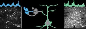
The activity of neuron types in the brain is imbalanced in mice with Huntington’s disease. The image depicts an example field-of-view from inhibitory (left) VIP (vasoactive intestinal peptide) neurons and excitatory (right) neurons recorded during behavior. Activity traces from a selected neuron for each type are shown above the images. [Sonja Blumenstock, Komiyama Lab, UC San Diego]

In particular, a class of inhibitory neurons known as vasoactive intestinal peptide (VIP) neurons, exhibited significantly reduced activity. VIP neuron activity is essential for normal learning, as these cells enable the brain to adapt and refine brain circuits during learning.

Reduced VIP neuron activity, the researchers reasoned, could be impairing the brain’s ability to function and learn properly. They sought to activate these cells to re-engage brain states that support learning. They tested this idea using optogenetics to stimulate VIP neurons.

“By activating the VIP inhibitory cell type, we gradually restored more normal activity patterns, and, very importantly, we also saw an improvement in the ability of the mouse to learn a motor task,” said Sonja Blumenstock, PhD, assistant project scientist at UC San Diego.

The results confirm VIP neurons as a key point of vulnerability in Huntington’s disease as well as a promising target for therapy. As to how this process works, the results suggest that modulating VIP neurons opens a “gate” that enables learning-related brain plasticity.

“This intervention restored more normal patterns of activity in the brain and improved movement in affected mice,” said Komiyama. “Importantly, the improvements persisted for days after stimulation ended, suggesting that the treatment triggered lasting beneficial changes in brain circuits rather than only temporary effects.”

The study provides important indications of where research could focus to normalize human brain function and facilitate brain recovery. Komiyama envisions a future scenario in which scientists could non-invasively activate the brain from outside the skull using novel approaches.

“Our study shows that despite the genetic defect, a precise intervention into the brain circuitry can lead to significant improvements in motor symptoms,” said Dudanova. “If we know which cells to target, we can retune the brain’s abnormal activity patterns. This gives hope for future therapies.”

The research also shows that corrections to specific brain circuit imbalances can restore function in a highly complex neurodegenerative condition, with similar potential in other disorders.

“We have come up with a way to allow the diseased brain to learn better,” said Komiyama. “The approach can improve behavior in diseased mice, and our hope is that a related approach will help people with impairment in their learning abilities.”

The post In Huntington’s Mouse, Optogenetic Activation of VIP Neurons Restores Brain Function appeared first on GEN – Genetic Engineering and Biotechnology News.

Biological Order Emerges from Tissue Boundaries, Drives Embryo Development

In a new study in Nature Materials titled, “Boundary geometry controls a topological defect transition that determines lumen nucleation in embryonic development,” researchers from European Molecular Biology Laboratory (EMBL) describe how interactions between tissue geometry impact development. 

In an early-stage mouse embryo, cells of the epiblast are polarized and give rise to all major tissues. The team investigated the fundamental principles governing the behavior of polarized cells that are present in bulk and the impact of physical constraints at tissue borders. By focusing on how cellular orientations influence each other and their environment, the researchers built a minimal model that predicts how organization changes when interactions are altered.

“For me, as a physicist, I may know why something works, but it’s still kind of magic to see that it’s all true in messy biological systems,” said Pamela Guruciaga, PhD, postdoctoral researcher at EMBL and co-first author of the study. “It was also super interesting coming from a pure physics perspective to come up with a common language to work with biologists.” 

In the cup-shaped epiblast, results showed different boundaries led to varying orientations for epiblast cells. When the boundary was lined with the extracellular matrix, the cells oriented perpendicularly. In contrast, when the epiblast was in direct contact with a neighboring tissue without a matrix, the cells aligned parallel to the boundary. The researchers found that the combination of these two orientations result in the appearance of structures, known as “topological defects.” 

“These are points in space where it is undefined in which direction an object should point,” explained Guruciaga. “For example, if a set of arrows is arranged in a starburst pattern, the center is a point where all directions are equivalent. These points are super relevant because they are very robust; you cannot easily destroy them.” 

To directly test whether the boundary shape controls the number of defects, the authors  altered the geometry of the epiblast. Perturbing embryo shape induced the formation of additional lumina at the predicted positions.  

“What I find most exciting is that these results identify a very general physical principle,” said Anna Erzberger, PhD, group leader at EMBL and co-corresponding author of the study. “We show that geometry alone can determine orientation patterns in three dimensions, independent of the microscopic details of the system. That means shape itself can act as a robust control parameter—not just in embryos, but across a wide range of biological and physical systems.” 

The post Biological Order Emerges from Tissue Boundaries, Drives Embryo Development appeared first on GEN – Genetic Engineering and Biotechnology News.

CAR T Cells Simultaneously Target Glioblastoma and Immune Cells 

Scientists have identified a new molecular target for CAR T-cell immunotherapy to attack both glioblastoma cells and tumor-supporting macrophages at once. A study published today in Nature shows promising preclinical results that could allow this approach to overcome the limitations of previous attempts to target glioblastoma with CAR T cells. 

“Our approach targets both the tumor and the environment that allows it to thrive,” said Sheila K. Singh, MD, PhD, professor of neuro-oncology and neurosurgery at King’s College London and McMaster University. “Instead of treating glioblastoma as only a mass of cancer cells, we need to think of it as a connected tumor-immune ecosystem. By going beyond the cancer cells alone, we are also targeting immune cells that help shield the tumor from treatment.”

Glioblastoma is an aggressive and lethal form of brain cancer where current treatments, including surgery, radiation and chemotherapy, only provide temporary benefits and are rarely able to prevent recurrence. Past attempts to develop CAR T therapies for glioblastoma have failed to produce sustained responses due to a number of challenges such as heterogeneous antigen expression, antigen loss, and microenvironmental barriers that treatments solely focusing on targeting the tumor cells have not been able to surmount. 

In particular, tumor-associated macrophages have been shown to be key contributors to glioblastoma progression. While macrophages normally play an important role in the immune response against infections, glioblastoma can recruit and reprogram these immune cells to promote tumor growth, suppress the immune system, and resist treatment. 

“CAR T therapy has been effective in some blood cancers, but translating that success to brain tumors has been difficult,” said Shan Grewal, an MD/PhD candidate at McMaster and co-lead author of the study. “Most approaches have focused on killing cancer cells alone. Our work suggests we may also need to dismantle the immune support system that helps glioblastoma survive.”

Using patient tumor samples, Singh’s team conducted multi-omic profiling studies that led to the identification of a promising target present both in glioblastoma cells and tumor-associated macrophages, called glycoprotein non-metastatic melanoma protein B (GPNMB). By engineering CAR T cells to target GPNMB, the researchers were able to attack glioblastoma tumors on two fronts and show potent antitumor activity in several preclinical models including patient-derived xenografts. 

While more work will be needed before this strategy can be evaluated in clinical trials, the study introduces a new framework to identify immunotherapy targets that could potentially be applied to a wide range of solid tumors beyond glioblastoma. 

Supporting this concept, a team at the University of Calgary has simultaneously published results in Nature Cancer from a first-in-human study using a similar approach in relapsed alveolar soft-part sarcoma (ASPS) and translocation renal cell carcinoma, two types of cancer that stably express GPNMB. In these patients, a CAR T-cell therapy directed against GPNMB was found to be safe and induced stable disease for up to three months, providing early clinical evidence supporting the feasibility of this therapeutic approach. 

The post CAR T Cells Simultaneously Target Glioblastoma and Immune Cells  appeared first on Inside Precision Medicine.

A Recap of the Inaugural Youth Mental Health Hub at SXSW London

In early June, SXSW London returned for its second year, gathering thousands of creatives, enthusiasts, entrepreneurs, and investors into the city to celebrate film, music, tech, and culture. As part of this year’s festival, the Child Mind Institute, in partnership with Wellcome, proudly presented the inaugural Youth Mental Health Hub – a week of programming dedicated to advancing solutions to one of the defining challenges of our time: the global youth mental health crisis. Through six thought-provoking sessions, leaders in clinical care, science, technology, policy, and media came together to explore how to strengthen prevention, improve early identification, reduce stigma, and build systems that meet young people where they are.

Here’s a look back at the inspiring conversations that took place throughout the week.

Beyond the Average: Understanding Vulnerability in the Digital Childhood Era

Beyond the Average: Understanding Vulnerability in the Digital Childhood Era

As artificial intelligence rapidly transforms the experience of childhood, experts explored how AI can both support and challenge young people’s mental health. The panelists discussed when and under what conditions young people may be most vulnerable as well as what systems we need to support them.

Moderator

Gary Wilson, Director of Research, Huo Family Foundation

Speakers
Catherine Sebastian, PhD, Head of Evidence for Mental Health, Wellcome
John Pickavance, PhD, Principal Data Scientist, Born in Bradford
Georgia Turner, Postdoctoral Research Associate, University of Cambridge
Michael Milham, MD, PhD, Chief Science Officer, Child Mind Institute

AI Is Already Shaping Childhood. Who Is Shaping AI? Balancing Innovation, Evidence, and Safety in Youth Mental Health

AI Is Already Shaping Childhood. Who Is Shaping AI? Balancing Innovation, Evidence, and Safety in Youth Mental Health

Youth are experiencing the impacts of AI earlier and more intensely than any previous generation has. This session explored the role of public leadership in anticipating harm before it becomes systemic — establishing guardrails, fostering digital resilience, and ensuring that innovation advances hand in hand with youth mental health and well-being.

Moderator
Sarah Aguiar-Borges, PhD, University of Cambridge

Speakers
Julia Gillard, former Prime Minister of Australia; Chair, Wellcome
Kanishka Narayan, UK Minister for AI and Online Safety
Giovanni Salum, MD, PhD, SVP, Global Programs, Child Mind Institute.

Youth Mental Health After Conflict: Healing, Resilience, and Rebuilding Systems

Experts shared insights on the unique mental health challenges facing children affected by war, displacement, and humanitarian crises. This session explored how societies can implement youth-centered systems grounded in prevention and use early identification to position youth mental health as a cornerstone of long-term recovery and resilience.

Moderator
Krupa Padhy, BBC Radio 4

Speakers
Dr. Mark Jordans, professor, Centre for Global Mental Health, King’s College London; Director of Research & Development, War Child
Emma Ferguson, mental health policy and advocacy specialist, UNICEF
Mohamed Ali, Director, Iftin Global

Dyslexia: Changing the Story

In a timely discussion, experts explored how dyslexia is currently understood in society, challenging current language and misperceptions that can impact a child’s confidence and mental health. Through a blend of personal experience and clinical expertise, the conversation focused on the need for evidence-based support and strengths-based approaches to help children and their families thrive.

Moderator
Kate Griggs, Founder, Made By Dyslexia

Speakers
Maggie Aderin, PhD, space scientist & educator; dyslexia advocate
Harold S. Koplewicz, MD, President and Medical Director, Child Mind Institute

Connection Continuum: Preventing Suicide and Combating Loneliness

Suicide is one of the leading causes of death among young people globally. This session gathered community, clinical, and digital leaders to explore what a more connected system of support looks like in practice. The panelists also discussed the important of recognizing warning signs, expanding access to evidence-based care, and prioritizing early intervention to help prevent youth suicide.

Moderator
Krupa Padhy, BBC Radio 4

Speakers
Victoria Hornby, CEO, Mental Health Innovations
Dean Perryman, Empty Chairs
Michael Milham, MD, PhD, Chief Science Officer, Child Mind Institute

Does Mental Health Science Funding Need a New Paradigm in the Age of AI?

With technology evolving faster than the science designed to understand it, experts examined how research, philanthropy, and clinical leaders can work together to build the evidence, safeguards, and infrastructure needed to protect children’s mental health in the digital age.

Moderator
Chelsea Clinton, Vice Chair, Clinton Global Initiative

Speakers
Miranda Wolpert, Director of Mental Health, Wellcome
Margaret Laws, President & CEO, HopeLab
Daria Bukhman, Co-Founder and Chair, Bukhman Philanthropies
Harold S. Koplewicz, MD, President & Medical Director, Child Mind Institute

The post A Recap of the Inaugural Youth Mental Health Hub at SXSW London appeared first on Child Mind Institute.

Mammographic AI Adds Predictive Power to Breast Cancer Risk Models

A study by Kaiser Permanente researchers has shown that integrating mammographic AI with polygenic risk scores and clinical risk models can improve breast cancer risk stratification, guiding both personalized breast cancer screening and chemoprevention.

The study, published in the Journal of the National Cancer Institute, is one of the largest and most diverse to evaluate the ability of the three approaches to predict breast cancer risk.

“Our goal is to improve our ability to assess a woman’s breast cancer risk so we can personalize breast cancer screening recommendations,” said lead author Vignesh Arasu, MD, PhD, a radiologist and research scientist at the Kaiser Permanente Division of Research. “Our study shows that each of the approaches identifies a distinct group of women, and that when all three risk tests are used, we increase our ability to differentiate high-risk and low-risk women and provide more personalized screening recommendations.”

The study included 82,957 women (75% non-Hispanic White, 9% Asian, 7% Latina, and 4% Black) who enrolled in the Kaiser Permanente Research Bank between 2003 to 2020. All the women had a recent negative mammogram, and none had previously been diagnosed with breast cancer or had a genetic mutation known to increase breast cancer risk.

The researchers calculated each woman’s breast cancer risk using three different approaches: The Mirai mammography AI risk score, which looks for risk-related imaging biomarkers; the Breast Cancer Surveillance Consortium version 3 clinical risk score, which considers factors such as age, race or ethnicity, family history of breast cancer, breast density, and body mass index; and the 313-SNP polygenic risk score (PRS) that assesses risk based on the presence or absence of 313 breast cancer-associated single nucleotide polymorphisms.

During 10 years of follow-up, 2471 (3%) women were diagnosed with invasive breast cancer or ductal carcinoma in situ.

Arasu and team report that the C-index for breast cancer prediction when all three risk scores were combined was 0.70.

“This means that if you take any women who actually will get breast cancer in the future and pass her information to a risk model, that model will say she has a higher risk about 70% of the time relative to women who won’t get cancer,” Arasu explained.

The C-index for the combined model was significantly higher than that for individual models with only the clinical risk score (0.62), which is used by most clinical practices, the PRS (0.61), or the Mirai score (0.66).

The C-index for the triple model was also significantly higher than that for a model that combined the clinical and the polygenic risk scores (0.66).

The increase in the C-index of 0.04 “represents a moderate but meaningful improvement in discrimination when incorporating all three risk domains compared with the clinical plus polygenic model alone,” Arasu told Inside Precision Medicine.

He added: “The C-index reflects overall model performance across all possible risk thresholds and is commonly used as an initial, global assessment when integrating new predictors, such as AI-derived measures, to gauge their added value. However, future work will focus on defining clinically actionable risk thresholds to better characterize how these improvements translate into real-world tradeoffs between benefit and harm.”

Importantly, the improvements in risk prediction were consistent over time and across four common self-reported racial/ethnic subgroups of Asian, Black, Latina, and White women, which the authors say indicates that incorporating mammographic AI and ancestry-adjusted PRS into clinical risk prediction models may benefit all women.

The study also found that among the women at highest risk for developing breast cancer, the clinical risk score alone identified 19% of the women who went on to develop breast cancer over a 10-year period while the combined model identified 26% of these women.

Although all three model types are becoming increasingly accessible and part of care, Arasu cautioned that “more research is needed to see the benefit of using all three scores before a program that uses all three should be implemented.”

He added: “We already are planning the next research step, which will be to study the combined model in the on-going national WISDOM clinical trial in the next 1-2 years. The WISDOM trial already uses a PRS and clinical risk score to assess risk. Now, we will be adding mammographic AI.”

“By identifying more accurately which women are truly at high-risk, we hope to find more breast cancers as early as possible, when they are most easily treatable,” said Arasu. “For women who are very high risk, there is also the potential to discuss, in addition to annual mammography, risk reduction with a medication, such as tamoxifen.”

The post Mammographic AI Adds Predictive Power to Breast Cancer Risk Models appeared first on Inside Precision Medicine.

Highly Sensitive ctDNA Test Improves Detection of Residual Pancreatic Cancer

A highly sensitive blood test that detects traces of tumor DNA in patients with localized pancreatic cancer identified substantially more patients with residual disease than conventional liquid biopsy testing, according to a prospective study published in Clinical Cancer Research. The findings suggest that more sensitive detection of circulating tumor DNA (ctDNA) could improve risk stratification after chemotherapy and surgery and help identify patients who remain at high risk for recurrence despite reassuring imaging results.

Researchers at Northwestern Medicine evaluated digital droplet polymerase chain reaction (ddPCR), a liquid biopsy approach that detects specific KRAS mutations, against standard next-generation sequencing (NGS), which surveys hundreds of cancer-associated genes but with lower sensitivity. Because KRAS mutations drive more than 90% of pancreatic cancers, the investigators hypothesized that focusing on this single, biologically important target would allow detection of extremely low levels of circulating tumor DNA that broader sequencing approaches often miss.

“We’re able to detect very high sensitivity in the blood for pancreas cancer,” said senior author Akhil Chawla, MD, clinical associate professor of surgery at Northwestern University Feinberg School of Medicine and a complex surgical oncologist at Northwestern Medicine. “We’re looking for extremely low levels of the DNA in the plasma.”

The prospective study followed 106 patients with localized pancreatic cancer from diagnosis through chemotherapy and surgical resection. Blood samples were collected before treatment, after chemotherapy, and following surgery to determine whether changes in KRAS ctDNA reflected treatment response and predicted patient outcomes.

At diagnosis, ddPCR detected tumor-derived KRAS DNA in 65% of patients, compared with just 17% using conventional NGS. The differences became even more striking after treatment. Following chemotherapy, ddPCR detected ctDNA in 60% of patients, while NGS detected it in only 5%. After surgery, ddPCR remained positive in 56% of patients compared with 9% using standard sequencing.

“What this publication is going to show is that yes, we can detect it with both, but we are missing a significant number of patients with standard sequencing,” Chawla said.

According to the study, patients whose disease was detected only by ddPCR represented a previously hidden intermediate-risk group. These patients had a median overall survival of 27 months after diagnosis, compared with 41 months among patients who tested negative by both assays. The findings suggest that standard liquid biopsy testing may underestimate the presence of minimal residual disease in many patients who appear to have responded well to therapy.

“We’re missing up to 60% of patients,” Chawla said. “Even at the time of diagnosis, and after treatment—particularly where we think it looks like on a CT scan after a patient’s undergone chemotherapy and had their surgical resection—everything looks great, and even the blood test that looks at ctDNA looks great. In sixty percent of patients we were still able to detect low levels.”

He adds, “Our goal is to get rid of that disease forever. Unfortunately, even with the work that we’ve done, we’ve shown that 60% to 70% of patients have recurrence of that disease after chemotherapy and surgery.”

The study also demonstrated that ctDNA dynamics over the course of treatment carried important prognostic information. Rather than relying solely on whether ctDNA was present or absent, investigators found that changes in ctDNA levels reflected treatment response.

“We’ve been able to see it even with chemotherapy and surgery,” Chawla said. “Patients that have that significant decline in the marker—not just the presence or absence, but even if they go from a high level to a medium to low level—those patients actually are benefiting from that treatment.”

Conversely, patients whose ctDNA levels remained stable or increased during treatment experienced substantially worse outcomes.

Unlike broad NGS panels, which search for hundreds of genetic alterations simultaneously, ddPCR focuses on a limited number of mutations with far greater analytical sensitivity. Chawla emphasized that the innovation was not a new laboratory technology but rather a new application of an established one.

“DDPCR can detect a single mutation with 1,000 times more sensitivity,” he said. “When we do that deep dive in pancreas cancer, what we’re really trying to do is identify whether this biomarker can, number one, be detectable in a high percentage of patients; number two, is it prognostic… and number three, can it really tell us how well a treatment is working?”

Because the assay targets the same KRAS mutations that are the focus of emerging targeted therapies, Chawla believes the approach could become increasingly valuable as those therapies enter clinical practice.

“I think there’s a lot of value in this test, particularly as we enter the age of KRAS-targeted treatments,” he said. “Our biomarker actually targets the exact same gene.”

Currently, surveillance after surgery relies primarily on CT imaging performed every few months, leaving clinicians with limited tools for detecting microscopic residual disease before recurrence becomes radiographically apparent.

“What the standard of care today is, is we just get CT scans every three months to give us an idea of when this comes back—we’re just kind of sitting on our hands and waiting,” Chawla said. “This blood test gives us an opportunity to be more active in that surveillance, potentially be more adaptive in our treatment.”


The post Highly Sensitive ctDNA Test Improves Detection of Residual Pancreatic Cancer appeared first on Inside Precision Medicine.

Stockholm3 Blood Test Detects More High-Risk Prostate Cancers Early 

A new blood test may help address one of the key challenges in prostate cancer detection—identifying aggressive forms of the disease early on. According to research led by a team at Karolinska Institutet, the Stockholm3 blood test detected more clinically significant cancer cases than the well-established, but problematic, Prostate Specific Antigen (PSA) screening test. 

The study appears in the Annals of Internal Medicine. Swedish researchers collaborated with teams from Europe and the U.S. on this work.

Prostate cancer is one of the top cancers among men globally, with an estimated 1.5 million new cases and 397,000 deaths annually. PSA testing has long been used for early detection. But, although PSA is prostate-specific, it is not cancer-specific. Elevated PSA levels can be caused by benign conditions as well as cancer and up to 50% of diagnosed aggressive prostate cancers are in men with low PSA values—below today’s cutoffs of PSA 3 ng/ml or PSA 4 ng/ml.

“There are a number of tests in development to improve on PSA alone. A lot of them, like Stockholm3, are fairly ingenuous and do a good job,” Mark Pomerantz told Inside Precision Medicine. He is a medical oncologist at the Dana-Farber Cancer Institute in Boston. Until one of these newer tests emerges as a winner, though, MRI is the gold standard for evaluating men with high PSA. “With MRI we can see the prostate in some detail,” Pomerantz said, “But it is expensive.”

The Karolinska-led researchers analyzed data from 12,670 men aged 50–74 from the population-based STHLM3-MRI study, which compared MRI-targeted and standard biopsy in men with elevated PSAs. In this more recent Annals study, the men were tested first with both PSA and Stockholm3 and followed for two years via national cancer registries, which allowed researchers to also identify cancer cases missed during the initial screening.

Stockholm3 detected 90 percent of aggressive cancer cases, compared to 74 percent for PSA.

“The test incorporates plasma protein biomarkers, genetic risk information from a polygenic risk score, and clinical factors such as age, family history, and prior biopsy history,” the study’s lead author Thorgerdur Palsdottir, told Inside Precision Medicine. Palsdottir is a researcher at the Department of Medical Epidemiology and Biostatistics, Karolinska Institutet. 

He added, “Together, these components provide a more comprehensive estimate of a risk of harboring clinically significant prostate cancer than PSA alone.” 

The components, he explained, map onto distinct biological axes rather than a single one. The kallikreins (PSA, free PSA, hK2) reflect prostate epithelial and tumor secretory activity, and the free-to-total PSA relationship helps separate benign enlargement from cancer. The polygenic score and family history capture inherited susceptibility—a man’s baseline predisposition—rather than anything about a tumor. GDF-15 is a stress-response marker that has been associated with more aggressive disease across several cancers. 

During the study follow-up, 443 men were diagnosed with clinically significant, i.e. aggressive, prostate cancer. Stockholm3 missed significantly fewer serious cancer cases than PSA, while the proportion of men incorrectly classified as high-risk was similar between the tests.

“These results point toward a potential change in how prostate cancer screening can be conducted. A more precise blood test could enable earlier detection of aggressive disease while reducing the number of unnecessary follow-up examinations and procedures,” said Palsdottir.

He adds that longer-term follow-up is needed to fully assess the effects on mortality and long-term outcomes. “The next important step is to evaluate longer-term outcomes, including disease progression, metastatic disease, and prostate cancer mortality.”

The post Stockholm3 Blood Test Detects More High-Risk Prostate Cancers Early  appeared first on Inside Precision Medicine.

Boys, Masculinity, and the Looksmaxxing Trend  

By now, you’ve probably heard of the term looksmaxxing. Think pieces about the trend have popped up all over the internet. And in a recent episode of Saturday Night Live, comedians poked fun at lookmaxxing influencers obsessed with having the perfect male physique.

While this new social media craze may seem silly, it’s impacting more boys than you might think. In a study conducted last year that surveyed over 3,000 young men (ages 16–25) from the United States, United Kingdom, and Australia, nearly two-thirds of participants were regularly engaging with masculinity influencers.

Teen boys are being encouraged to change the way they look in order to fit a certain standard of attraction. The growing amount of looksmaxxing content they see online can have real effects on their self-esteem and mental health.   

What is looksmaxxing?  

Looksmaxxing originated nearly a decade ago in incel forums where men blamed their lack of romantic partners on the belief that female sexual selection is primarily based on physical qualities. So men who aren’t born with traits desirable to women are doomed to fail romantically. While traditional incels wallow in this fate, looksmaxxers seek to enhance their appearance to become more attractive. Their community claims that there is a universal standard for what the ideal man (and woman) should look like.

This is determined by a rating system called the PSL scale — the name being an amalgamation of three prominent misogynistic incel forums of the 2010s. There are many factors that go into the scaling, such as eye shape, jaw size, nose angle, and body fat percentage. Along this scale, you can land in four categories: subhuman, normie, Chadlite, and Chad (the ultimate catch).

During the pandemic, looksmaxxing went mainstream, merging with “manosphere” content on social media platforms like TikTok and Instagram. The trend became less about the ability to attract women and more of a competition among boys and men as they engaged in mog-offs — online contests where people have their faces analyzed and compared by facial recognition software to determine who’s better looking.

Self-improvement practices have gained popularity among boys. Some are considered to be softmaxxing, like developing skincare routines or eating high-protein diets, and others to be hardmaxxing, like using growth hormones or getting cosmetic surgery.

Prominent young influencers like Clavicular represent the extreme side of looksmaxxing. He practices bonesmashing (using a hammer on facial bones to try to form more angular features), injects himself with testosterone, and takes meth to maintain a low body fat percentage while still having a muscular physique.

Looksmaxxing and new beauty standards

The rise of looksmaxxing seems to have a caused a ripple effect among teen boys. While the ideal look has centered on big muscles and washboard abs for decades, there’s now an added pressure on facial beauty that’s typically been reserved for girls.

“With some of the teen boys I work with, most of whom already have self-esteem issues, I think there is a lot more concern about how they look,” observes Alnardo Martinez, LMHC, director of the Pediatric OCD Intensive Program and a mental health counselor at the Child Mind Institute. “They want to have the strong jaw, really big muscles, clear skin, and a perfect haircut.”

However, Martinez notes that it sometimes take a while for boys  to admit that they feel this pressure. They may insist that they don’t really care about that stuff. “But then, maybe a few months later, it comes out that there is a lot of comparison. They’re spending a lot of time in front of the mirror or in the bathroom trying to create this perfect image,” he observes.

What teen boys think about looksmaxxing and self-improvement

We talked to young men who were critical of Clavicular and the impact looksmaxxing can have on teens but were positive about engaging in some form of physical self-improvement.

Wyatt, now 19, remembers comparing his jawline to his peers’ when he was in 7th grade. “I just felt like they had really sharp jawlines. And I was just like, ‘Oh, I want to get closer to that.’” He would also come across TikToks advertising rubber chewing blocks and chin exercises meant to strengthen the jawline.

And so, Wyatt began to do jaw exercises he’d found online, reciting the alphabet while stretching out the muscles. “I would go through my Zoom classes throughout the day and then after that was done, I’d just go into the bathroom and go through the whole exercise. It would take like an hour sometimes,” he recalls. “It turned into more like a self-care, self-improvement session. I would do that every day after my classes. I didn’t feel like I was done with school until I finished my jawline routine.” He took photos to document his progress.  

Wyatt feels like the routine had a positive effect, because he was able to see an improvement. “I felt more satisfied with myself, a little more confident.”

Lev, now 19, remembers wanting to have some control over his body when going through puberty in high school. “Puberty is not a straightforward process. It’s not all peaches and cream. Your body changes, and it can be uncomfortable,” he explains. “But with lifting and strength training, it was very exciting to see this, you know, man energy that came out of it. I wanted to harness that and really take it by the reins. Have some agency as a man.”

And while he rejects the extreme parts of looksmaxxing, Lev does regularly practice self-improvement through weight lifting, skin care routines, and taking GLP-1 weight loss medication.

How looksmaxxing can impact boys’ mental health

Since looksmaxxing places such a strong emphasis on achieving a very specific look, clinicians are concerned about its influence on teens. “Self-esteem is pretty fragile during puberty,” Martinez says. “There’s already a ton of comparison and perceived flaws that teens don’t love about themselves.”

These insecurities can be exacerbated by the type of content teens engage with online, Martinez explains. Along with ChatGPT bots specifically designed to judge aesthetics, Reddit threads such as r/Mewing and websites like Looksmaxxing Forum encourage boys to post pictures of their faces and bodies to get rated by their peers. Boys as young as 13 visit these forums, posting pictures and asking for tips on how to improve their looks.

“These are generally places where people are already pretty harsh and critical. These boys are receiving a lot more ‘confirmation’ around the perceived things that are wrong with them or that they need to change,” Martinez says. “And it just feeds into the already present negative self-image and self-talk.”

He explains that this type of social media engagement can also compound underlying mental health issues like depression and social anxiety. “They might be less likely to go out and talk to people because they’re thinking, ‘Everyone is going to see this one thing that everyone else has told me is wrong with me. So now I can’t go out,’”he says.

Martinez is also concerned that online content can negatively affect teens with body dysmorphic disorder (BDD). “If they think they have a big nose, for example, they might go on these Reddits and ask, ‘What does my nose look like? Is it too big?’ There are trolls out there. Someone is going to say yes and then that’s going to make the BDD symptoms even worse.”

When behaviors might be concerning

In some ways, teen boys taking part in more self-improvement practices could be seen as a good thing. They’re exercising, taking care of their skin, and eating more balanced diets. The issues begin when these types of practices turn into obsession. And given the underlying ideology of looksmaxxing and the nature of social media, things can become unhealthy.

According to Martinez, there are some changes in behavior to look out for that indicate you might want to step in.

One clear change, he says, is a noticeable shift in the amount of time they’re spending on grooming themselves. “Maybe they were someone who would typically just get up and run out the door without washing their face,” he says. “But now they’re spending a lot more time in the bathroom and asking a lot of questions about how they look.”

Another warning sign can be a big change in personality. “Irritability is a big one that we’ll see a lot,” he says. “They’re unhappy with how they look, so this increases a general level of irritation.”

These behaviors paired with an unusual uptick in time spent on social media, Martinez explains, can be a sign that something’s wrong and support is needed.

How to support your child

If you’re worried that your child might be engaging in looksmaxxing-related behaviors to an unhealthy degree, says Martinez, there are a few things you can do:

  • Open communication. Martinez suggests approaching your child with curiosity. “You could start the conversation by saying something like, ‘So have you heard about this? What do you think about it? Have you ever had any thoughts yourself about how you look or desires to change your body or face?’ And then give them some space to be open and vulnerable about it. Validate their experience.” 
  • Find out where your child is getting their information. “Read it together, talk about it, and see what your child thinks about it,” Martinez advises. “And if it’s promoting something dangerous, then you can talk to them about how those practices can be harmful and what could actually happen if they do some of those things.”
  • Encourage male role models. “There’s a patient I work with now who doesn’t have a present dad,” Martinez explains. “His mom tries to talk to him about things like body image, but he feels like she doesn’t understand and can’t relate. So having someone that he can talk to and be open about this stuff with, especially someone who can also share their own struggles, can be really helpful.”
  • Seek help from a mental health professional. This is especially important if you find out that your child has been engaging in extreme forms of looksmaxxing such as bonesmashing or starvemaxxing. Martinez recommends looking for a clinician who specializes in body image or body dysmorphic disorder.

A lot of parenting comes down to open communication around what your kids are seeing and what they’re feeling. We all have things about our bodies that we might not like and wish we could change, says Martinez, and it can help to normalize those feelings. “And then you can discuss how they can make changes in healthy ways,” he suggests. “Go over what’s a realistic change and what’s a dangerous change.”

The post Boys, Masculinity, and the Looksmaxxing Trend   appeared first on Child Mind Institute.

Digitize or Fall Behind

Bioprocessing companies risk slowing scientific progress unless they embrace digital-data capture and greater collaboration, according to Alexander Seyf, CEO of Autolomous, a company developing digital manufacturing solutions for cell and gene therapies.

Speaking about the industry’s biggest challenges, Seyf describes poor data management as the “elephant in the room,” arguing that too much crucial information remains trapped in paper records, spreadsheets, and isolated systems.

“Everybody wants to have AI,” Seyf says. “But where do you have your data? If it’s in binders, there’s not much you can do.”

According to Seyf, the path toward more efficient manufacturing, stronger clinical outcomes, and meaningful AI applications begins with digitizing information from the earliest stages of research. He believes many organizations make the mistake of waiting until their science is mature before investing in digital infrastructure. “The sooner you start, the better it is,” he says. “Pen and paper do not prevail, and pen and paper do not transfer.”

Seyf argues that the consequences extend far beyond operational inefficiencies. When data remain inaccessible or fragmented, researchers lose opportunities to learn from past experiments, identify patterns, and accelerate scientific discovery. He stresses that the industry must become more willing to share non-commercially sensitive knowledge, particularly in areas such as rare diseases and advanced therapies, where patient populations are limited. “We are all here to serve patients,” he says. “Protect your intellectual property, but also share the learnings.”

One of his strongest criticisms is directed at the scientific community’s tendency to focus almost exclusively on successful outcomes. Seyf believes failed studies and unsuccessful trials often contain lessons that could prevent others from repeating the same mistakes. “A lot of publications want to publicize only the good news,” he says. “That’s fundamentally wrong. We need to learn from failures.”

To illustrate his point, Seyf compares the biotechnology sector with the aviation industry. Modern airlines routinely share information about incidents and technical problems to prevent future accidents, creating a culture of collective learning and safety. “If something goes wrong, everybody in the world knows about it and knows how it was managed,” he says. “We are also dealing with people’s lives. The only way for us to improve is to share.”

Seyf also highlights the growing role of AI in healthcare. Although consumer AI systems have benefited from vast amounts of publicly available information, healthcare still operates with a relatively small pool of accessible data, he says. Expanding that foundation, he argues, could unlock major advances in diagnosis, drug development, and personalized medicine. “Imagine what we could do,” he says. “The progression of science is unlimited.”

For commercial bioprocessors, his recommendation is straightforward: digitize from day one. Capturing research, development, manufacturing, and clinical data in digital formats not only improves collaboration but also preserves institutional knowledge when employees move on. “Every time a scientist leaves, the knowledge goes with them,” Seyf says. “But when it is digital, the knowledge stays with the company.”

As cell and gene therapies continue to evolve, Seyf believes the industry faces a choice. It can continue operating in silos, or it can embrace transparency, digitalization, and collaboration to speed innovation and deliver better outcomes for patients. “The reason humanity has progressed,” he says, “is because we shared.”

The post Digitize or Fall Behind appeared first on GEN – Genetic Engineering and Biotechnology News.