

Background: The rapid integration of artificial intelligence (AI) and medical big data into health care is transforming diagnosis, treatment planning, and research. However, formal education in these areas remains limited in undergraduate medical curricula, particularly in China. Objective: This study aimed to investigate clinical medicine undergraduates’ familiarity with AI and medical big data, their perceived need for related courses, and their preferred curriculum design and assessment methods. Methods: A cross-sectional, web-based survey was conducted at Zunyi Medical University, Guizhou, China, from January 10 to 17, 2025. In the institutional context of this study, “clinical medicine” included related clinical-track specialties such as pediatrics and psychiatry. All eligible students (N=1094) were invited, and 871 (79.6%) were included in the final analysis. The self-administered questionnaire was developed based on a literature review and expert consultation, with content validity quantified using the content validity index. Descriptive statistics were used to summarize response distributions. For ordinal outcomes (items 1-14), adjusted ordinal logistic regression models were applied, with gender and grade as predictors and major as a covariate. Given the small number of third- and fourth-year students, grade was modeled as an ordered trend variable. For nominal outcomes (items 15-16), group differences were assessed using chi-square tests or Fisher exact tests, as appropriate. Results: A total of 871 students were analyzed, of whom 62.6% (n=545) were women. Overall familiarity with AI and medical big data was limited: 34.8% (303/871) agreed or strongly agreed that they were familiar with the topic, and only 33% (287/871) reported having at least some prior learning experience. In contrast, the perceived educational need was high: 94% (819/871) considered such a course at least somewhat necessary, 57% (497/871) reported that the course was needed or very needed, 75.5% (658/871) indicated that they would likely or definitely enroll, and 56.5% (492/871) reported that they would likely or definitely engage in self-directed learning. Personalized teaching based on textbooks (566/871, 65%) or open-book examinations (633/871, 72.7%) was the most preferred instructional and assessment format. Preferences for course materials and assessment methods differed by grade but not by gender. Conclusions: Early-stage clinical medicine undergraduates demonstrated limited familiarity with AI and medical big data but expressed a strong demand for related education. Students preferred structured yet flexible instructional formats and open-book assessments. Although the findings are based predominantly on first- and second-year students, they support the development of staged, practice-oriented AI and medical big data curricula tailored to the needs of early-stage clinical medicine undergraduates.
<img src="https://jmir-production.s3.us-east-2.amazonaws.com/thumbs/a22edb5d5bd9dfd0ef9d0e91d5560eaf" />
Peptistar Began Operation of the Asahi Kasei FO-MD System at Manufacturing Scale
Japanese CDMO Peptistar reports that it has integrated Asahi Kasei’s forward osmosis–membrane distillation (FO–MD) system into its facility for trial production of active pharmaceutical ingredients (APIs).
Asahi Kasei announced in 2018 the development of a system that dehydrates and concentrates liquids without the application of heat or pressure. This reduces the number of freeze-drying batches and the amount of time required for freeze-drying, thereby shortening API manufacturing time. Peptistar has begun operation of the system at manufacturing scale as part of its evaluation toward GMP production.
![FOMD-system-for-concentration-without-heating-or-pressurization-installed-at-Peptistars-peptide-and-oligonucleotide-API-manufacturing-facility [Asahi Kasei]](https://www.genengnews.com/wp-content/uploads/2026/07/FOMD-system-for-concentration-without-heating-or-pressurization-installed-at-Peptistars-peptide-and-oligonucleotide-API-manufacturing-facility-300x225.jpg)
Recently, demand for APIs has shifted from traditional, high-volume small molecules to a broader need across biologics, peptides, oligonucleotides, viral vectors, and more, according to officials at both companies. API needs are becoming increasingly complex due to their high specificity and growing role in next-generation therapeutics.
Some of the next-generation APIs such as peptides and oligonucleotides are heat sensitive. Their manufacturing processes have thus relied on the costly, time-consuming, and energy-intensive freeze-drying method, which can remove solvents without heating, to obtain APIs with high quality explains an Asahi spokesperson.
Although the freeze-drying process can be shortened by concentrating the raw material solution to reduce the volume of liquid feed prior to the freeze-drying step, conventional concentration technologies such as vacuum distillation carry the risk of quality degradation due to heating, and the formation of precipitates caused by changes in solvent composition during the concentration step, adds the spokesperson.
![Overview-of-the-FOMD-system-for-concentration-without-heating-or-pressurization. [Asahi Kasei]](https://www.genengnews.com/wp-content/uploads/2026/07/Overview-of-the-FOMD-system-for-concentration-without-heating-or-pressurization-731x1024.jpg)
Asahi Kasei’s system for forward osmosis (FO) and membrane distillation (MD) addresses such manufacturing challenges by concentrating the raw material solution for pharmaceutical applications without applying heat or pressure, notes another Asahi official, explaining that FO utilizes an osmotic pressure difference across a membrane to remove water from liquids, achieving highly concentrated API solutions under mild conditions. MD leverages a vapor pressure difference across a membrane to remove volatile components such as acetonitrile, alcohol, or ammonia, at or below room temperature.
Asahi Kasei says it looks forward to studying the prospects for future commercialization of the FO–MD system.
The post Peptistar Began Operation of the Asahi Kasei FO-MD System at Manufacturing Scale appeared first on GEN – Genetic Engineering and Biotechnology News.
Why California’s carbon manure math doesn’t add up
Something stinks in California’s climate policies.
Years ago, the state set up a system that pays cattle farmers across the country to turn the methane emitted from cattle manure into natural gas, encouraging the dairy sector to produce a gas we burn instead of one that just pollutes the air.
It’s become wildly popular because the subsidies are extremely lucrative. But a growing body of research suggests the program is a case study in the shortcomings of our preferred approaches to climate action. Instead of simply forcing industries to directly cut their pollution or pay for it as a cost of doing business, legislators have repeatedly opted to set up convoluted incentive systems that swap climate responsibilities between parties and regions. As studies have shown again and again, these carbon offsetting and trading schemes often dramatically overstate the emissions reductions actually achieved in the one place that matters: the atmosphere.
The dairy program illustrates a particular version of this problem, muddling the impacts of different types of greenhouse gases in a way that researchers argue will lock in more warming in the future.
Despite this and other concerns, California regulators decided in 2024 to extend parts of the program beyond 2050. And a recent proposal by the state’s air resources board could send millions of additional dollars to dairy farmers as part of a plan that would ease restrictions on major greenhouse-gas producers.
Here’s how the system works: The state’s climate regulations require the transportation fuels industry to lower the carbon dioxide levels in its products over time—or purchase credits from other parties that cut fuel emissions, including cattle farmers.
Dairies generally spray cattle manure into giant open lagoons, where microbes gobble up organic matter and produce methane as a by-product. But if farmers set up what are known as anaerobic digesters, the sludge is redirected into covered vessels that capture the biogas, which can be converted into natural gas and injected into a pipeline. It can then be used to fuel certain vehicles or generate electricity in a power plant. Either way, petroleum companies can pay those farmers for Low Carbon Fuel Standard (LCFS) credits, to meet regulatory requirements in lieu of reducing the emissions from their own fuels.
Burning biogas in a bus or turbine still releases carbon dioxide, but the idea is that this process reduces market demand to extract natural gas from the ground and avoids the release of methane, which is a far more powerful greenhouse gas (at least initially). In fact, methane is so much more powerful that under California’s program, “adding one average biogas-powered vehicle to the fleet would produce enough LCFS credits to cover the deficits incurred by 26 similar gasoline-powered vehicles,” according to Aaron Smith, a UC Berkeley economist.
But there’s a problem with this carbon math. California assumes that methane exerts about 25 times the warming effect of carbon dioxide over a 100-year period. That’s not how it really works in the atmosphere, though.
Methane is very powerful, but it also breaks down quickly, generally within a couple of decades. Meanwhile, carbon dioxide builds up cumulatively in the atmosphere—and much of whatever we emit will continue heating up the planet for hundreds to thousands of years.
So, in effect, the state has created a system that reduces short-term warming at the cost of increasing all-but-permanent warming. Any methane that digesters capture today would have caused extra-powerful warning if released, but by 2050 that effect would have mostly faded away. Meanwhile, that additional carbon dioxide we permitted in its place could continue warming the world for millennia.
It is a good idea to cut methane emissions, and dairy digesters achieve this (though not always as effectively as hoped). But we can’t swap a decrease in short-lived greenhouse gases for an increase in long-lived ones if we hope to keep global temperatures within relatively safe levels in the coming century, as researchers have long warned. We have to slash both.
The problem I keep returning to, after years of covering carbon markets and offsets, is this: We need to clean up every sector, completely, over the next few decades. It’s increasingly untenable for so many of our climate ambitions to turn on getting one industry to make progress on paper by paying another one to reduce emissions, at a point when every business in every industry needs to be racing toward net zero.
It’s time to move past the idea that we need to reward sectors for doing us the favor of not polluting the atmosphere, and simply require them to stop unloading the huge environmental burden of their business onto society.
This article is from The Spark, MIT Technology Review’s weekly climate newsletter. To receive it in your inbox every Wednesday, sign up here.
Sheffield first council to oppose NHS FDP Palantir contract
Sheffield City Council has become the first local authority to approve a motion formally opposing Palantir’s involvement in the NHS FDP.
Effectiveness of the Group Unified Protocol for Emotional Disorders in Adults in a Public Mental Health Setting
Conditions: Emotional Disorders; ANXIETY DISORDERS (or Anxiety and Phobic Neuroses); Depressive Disorder; Somatic Disorders
Interventions: Behavioral: UP; Behavioral: TAU
Sponsors: Patricia Fernández Couto; University of La Rioja
Recruiting
Interventions: Behavioral: UP; Behavioral: TAU
Sponsors: Patricia Fernández Couto; University of La Rioja
Recruiting
<![CDATA[Phase 3b trial shows centanafadine eases adult ADHD and anxiety symptoms fast, with expected safety profile, as FDA review nears.]]>
Biological aging might help to explain the rising risk of early-onset cancer
Nature Medicine, Published online: 01 July 2026; doi:10.1038/s41591-026-04517-0
Cancers have risen rapidly in younger adults in recent generations. Analyzing large population-based cohorts, we found that recent generations exhibited signs of faster biological aging than older ones, and that systemic and organ-specific aging markers were associated with an increased risk of early-onset solid cancers.
Author Correction: Spinal cord Tau pathology induces tactile deficits and cognitive impairment in Alzheimer’s disease via dysregulation of CCK neurons
Nature Neuroscience, Published online: 01 July 2026; doi:10.1038/s41593-026-02356-3
Author Correction: Spinal cord Tau pathology induces tactile deficits and cognitive impairment in Alzheimer’s disease via dysregulation of CCK neurons
First 3D Structure of Malaria’s “Moving Junction” Solves Infection Mystery
For nearly half a century, scientists have known that malaria parasites force their way into human red blood cells (RBCs) through a ring-shaped structure called the moving junction (MJ). What no one could work out was what it actually does. The structure assembles, does its job, and dissipates in the space of 60 seconds—gone before anyone can get a close look.
A team at Columbia University has now finally caught the moving junction in the act. By freezing parasites at the onset of invasion and lifting the intact complex straight out of the cell, the researchers obtained the first high-resolution view of its three-dimensional structure. What they saw overturned a decades-old assumption about how the parasite gets in. Rather than a passive doorway, the moving junction turns out to be a molecular machine that actively remodels the host cell’s membrane to help the parasite force its way inside.
The findings detail how the team obtained the structure and then used it as a blueprint to design a mini-protein, from scratch, that blocks invasion—a proof of concept for a new kind of antimalarial drug.
“We’ve known for decades that this structure is essential for the parasite to get into a cell, but not how it actually works,” said Chi-Min Ho, PhD, an assistant professor in the Department of Microbiology and Immunology at Columbia University Vagelos College of Physicians and Surgeons and the study’s senior author. “Pulling it directly out of the parasite intact let us finally ask that question directly.”
Ho is senior author of the team’s published paper in Cell, titled “Structural basis for host membrane binding and remodeling by invading malaria parasites.” In their paper, the team stated in summary, “This work represents a major step toward resolving the decades-long mystery surrounding the structure and function of the malarial MJ, underscoring the power of pursuing native structures and laying the foundation for structure-guided design of next-generation antimalarials.”
Malaria still kills roughly 600,000 people a year, the overwhelming majority of them young children in sub-Saharan Africa, and the parasite is steadily becoming resistant to frontline drugs. “Malaria morbidity and mortality are directly linked to the invasion and replication of the malaria parasite Plasmodium falciparum in human red blood cells (RBCs),” the authors wrote. The malaria parasite life cycle involves two hosts, humans and Anopheles mosquitoes, and infecting human RBCs and hepatocytes, as well as mosquito salivary glands.
The disease starts with a single event: a parasite breaking into a red blood cell. “Parasites establish infection by invading host cells in a rapid and precisely choreographed process …” the team continued. In an infected person, trillions of parasites are released and invade every 48 hours in synchronized waves. This rhythmic cycle of rupture and reinvasion drives the periodic fevers malaria is known for. “After gliding, reorientation, and initial attachment, parasite internalization is initiated by the formation of a ring-shaped ultrastructure called the moving junction (MJ), which anchors the parasite to the host cell,” the researchers explained.
The same moving junction machinery is used across every species and every stage of the parasite’s life cycle, which has made it one of the most sought-after targets in malaria research. For antimalarial drug and vaccine development, block it, and you stop infection at its source.
The moving junction has been a puzzle since 1978, when scientists first observed in electron microscopy images a mysterious thickening of the membrane where parasite meets cell. Researchers eventually identified the four parasite proteins—AMA1, RON2, RON4, and RON5—that assemble into the junction’s basic building block, and confirmed that all were essential for invasion. But what the structure actually did remained unknown, because it survives for a minute or so and refuses to reassemble in a test tube. “Efforts to address this critical gap in understanding have been thwarted by the short-lived (60–90s) nature of the complex, as well as by the difficulty of recapitulating it in heterologous systems for detailed biochemical and structural study,” the researchers stated.
The Columbia team got around this by stopping invasion mid-stride. Using a compound that halts the parasite’s internal motor without preventing the junction from forming, they stalled parasites partway into red blood cells, then extracted the fully assembled AMA1-RON complex—the building block from which the whole junction is constructed—and imaged it with cryo-electron microscopy (cryo-EM), a technique where molecules are flash-frozen and imaged with an electron beam at extremely high magnifications to reveal their shape in atomic detail. The result was a sharp, three-dimensional view of that building block. The researchers noted that it was quite strikingly shaped like a sailboat, with the AMA1 protein forming a “sail” above the cell surface and the three RON proteins forming a broad “hull” pressed against the membrane below.
The biggest surprise was in the hull, where the team found clues that finally hinted at the moving junction’s role in invasion. The face of the structure pressed against the host membrane is blanketed with positively charged anchors, and the surface is studded with short helices that drive deep into the membrane like wedges. “These short helices insert asymmetrically into one leaflet of the membrane, displacing lipid headgroups and applying lateral pressure to generate local membrane deformations.”
Both features are widely recognized hallmarks of a well-known family of cellular machines that bend and reshape membranes. Their structural findings, they noted in their report, reveal “a highly unusual molecular staple that exhibits the hallmarks of a powerful membrane-remodelling machine.”
To test whether the structure could indeed deform a membrane, the researchers synthesized the parasite’s wedge-like helices and added them to artificial membrane bubbles. The membranes thinned and punctured. Meanwhile, weakened versions of the helices left the bubbles intact. The team concluded that the moving junction appears to pull the host membrane into shape, likely working in concert with the parasite’s motor to lever the parasite inside.
“It had been pictured as a kind of series of staples or spot-welds, making up a passive ring the parasite hauls itself through,” said Meseret Haile, the study’s first author and a PhD candidate in Ho’s lab. “What we see instead is a machine built to reshape the host cell’s own membrane. That changes how we think about the whole event.” In their paper, the team added, “Our work reveals that, although visually suggestive of canonical tight junctions, the MJ differs fundamentally in function, serving as a dynamic portal that orchestrates parasite internalization, rather than a static adhesion molecule.”
Beyond finally revealing how the moving junction allows the parasite to invade, the structure also gave the team a precise map of where and how AMA1 grips its partner protein, the contact that holds the entire junction together. Using a machine learning-powered protein-design tool together with their structural information, the researchers designed a mini-protein to break that grip. Their best candidate blocked parasites from invading red blood cells in a dose-dependent way and left already-infected cells unaffected, confirming that it works specifically by stopping entry rather than through general toxicity.
The designed mini-protein is a first proof of concept, not a drug, and will need considerable refinement before it could be tested in people. But it demonstrates an exciting new strategy: using near-native structures to design invasion-blocking mini-proteins against a target that has long frustrated conventional approaches. The same structure also clarifies how several leading anti-malaria antibodies work, information that could feed back into vaccine design. “Our successful proof of principle demonstrates the potential power of context-driven binder design for challenging systems, offering a previously unexplored avenue for therapeutic intervention,” they wrote. “In addition to their therapeutic potential, these binders may also serve as powerful tools for probing the functional relevance of specific protein interactions.”
Daphne Kaxiras, an MD-PhD student in Ho’s lab who led the inhibitor design, said, “Once we could see the target in its real setting, designing something to block it became a tractable problem. That’s the part we’re most eager to build on.”
The team’s approach, imaging fragile complexes captured directly from the organism and using them to guide design, may apply to many other parasites and pathogens that are notoriously difficult to study.
The post First 3D Structure of Malaria’s “Moving Junction” Solves Infection Mystery appeared first on GEN – Genetic Engineering and Biotechnology News.
The Microbiome’s Growing Role in Cancer Immunotherapy
Much of cancer research and therapy focuses on the direct impacts on cancer cells. However, understanding the broader context of cancer as a component of a patient, rather than an isolated invader, has opened a variety of insights and treatments for patients with cancer. Investigations of how the microbiome impacts cancer and immunotherapy was the prime focus of the second session on June 23, 2026 at the Frontiers in Cancer Immunotherapy Symposium hosted by The New York Academy of Sciences.
GVHD and the microbiome
Opening the discussion, Marcel van den Brink, MD, PhD, president of City of Hope Los Angeles and City of Hope Medical Center spoke about the role of the intestinal microbiome in cancer immunotherapy.
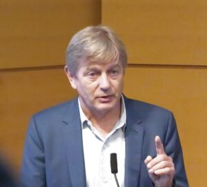
He began by describing the history of graft versus host disease (GVHD), pointing out that while early work from the 1970s suggested that germ free mice had reduced instance of GVHD following transplants, more current work has pointed to the intestinal microbiome as an immune system modulator.
“Protection of the commensal anaerobes is beneficial,” he said. He explained that Enterococcus has a habit of dominating a population with reduced diversity, pointing out that it “happens very frequently within the context of allogenic transplant, and again is linked with graft versus host [disease].”
“So we try to understand how that happens, why do you get that domination?” van den Brink described how damage to the internal lining of the gut by chemotherapy or XRT conditioning can lead to alloreactivity of immune cells. Damaged enterocytes are less able to produce lactase, leading to increase in lactose availability, which can help drive the growth of Enterococcus species, including E. faecillis—a primary species found in patients who develop GVHD. Concurrently, bile acids can have an immune suppressive effect.
He went on to share results of two published studies exploring the role of immune cells in this cycle and potential interventions. He summarized this work saying, “The protection of the commensal anaerobes is critical.
“That’s probably the easiest point that I can make, if you think about using the gut microbiome as a target to improve outcomes for cancer patients.”
As the gut is a complicated ecosystem, and there are many angles of research, the van den Brink lab is also now exploring other avenues of research that do not involve antibiotics in addition to their work with antibiotics and other therapies.
Probiotic engineering
The second talk in this session, presented by Nicholas Arpaia, PhD, associate professor of microbiology and immunology at Columbia University, explored the possibility of personalized cancer immunotherapy with the use of engineered probiotics.
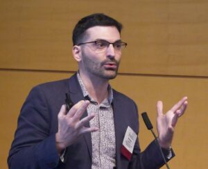
His work has focused on exploring the interactions between bacteria and the tumor microenvironment (TME) and how bacteria can act as a sort of Trojan horse to access the inner tumor environment.
In terms of cancer immunotherapy, Arpaia began by saying, “hopefully I’ll be able to convince you that utilizing bacteria is a potential path forward.” Bacteria, he argues, have a bright future in the field with a strong and growing research background based on the publications, companies formed, and clinical trials over the last 20 to 30 years.
He continued describing the wide scope of the field, both in how bacteria are engineered, and in how those bacteria are delivered. While the immune-oncology space has tended towards engineering payloads that modify the TME or deliver neoantigens, there are other approaches aimed at delivering toxins or modifying the metabolism within the TME. Further, Arpaia shared details on the differences between intravenous (IV), intratumor, or oral delivery. He pointed out that bacteria injected intravenously have been found in the cores of tumors. “It’s been speculated that this occurs because of the amenable conditions within the tumor.”
The question then arises, how can this behavior be beneficial to cancer therapy? “Features of bacteria themselves can activate the innate immune system,” Arpaia said. “If you then couple that with something that’s going to help activate the adaptive immune system, it gives us all the signals we need to really get long-term durable and effective responses.”
While many bacterial strategies involve the bacteria bringing specific payloads to the TME, much of his work explores a strategy of quorum-based lysis or a synchronized lysing circuit.
“Essentially what we should observe is that there’s growth, they hit a quorum threshold, so this synchronized lysis event occurs, a few of the bacteria remain, and the entire population undergoes these cyclic events again.”
Following the lysis event, what remains is “just a massive bag of innate immune stimulatory ligands.” The payload is released over and over through this synchronized lysis of the bacteria. Arpaia summed the process: “They grow, they undergo a lytic event, they grow back, and the entire process again occurs again and again.”
Tumor-associated bacteria in space
The final talk seamlessly transitioned from the discussion of bacterial lysis deep within the tumor to a discussion on how the location of tumor-associated bacteria within the TME can impact therapy approaches.
Susan Bullman, PhD, associate professor of immunology at the University of Texas MD Anderson Cancer Center began her discussion by taking a step back from cancer. “What I’m going to talk about is the native colonization of tumors by bacteria, by members of our microbiome,” she said.
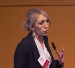
She explained that her group is “particularly interested in oral gastrointestinal cancers and understanding how microbes disseminate from our microbiome and can infiltrate human tumors to modulate the TME.”
Bullman described how certain bacterial that or typically restricted to the oral cavity can migrate and infiltrate cancers throughout the gastrointestinal (GI) tract. She focused specifically on Fusobacterium nucleatum, which not only has been consistently identified in GI tract tumors, but has also been found to negatively impact patient outcomes.
“When this microbe is enriched in the tumor, patients tend to have an increased risk for relapse, metastases and overall poor prognosis,” she said. Bullman explained that there is variability between tumor types and likelihood of tumor infiltration by microbes—with GI tract tumors having a higher instance of bacterial infection. Further, there is a heterogenous distribution of the bacteria within the tumor itself and while this bacterium is not the only microbe within the tumor tissue, her work aims to understand how this species modulates the TME.
She asked, “When these microbes get into a tumor tissue or infiltrates the tumor tissue, what exactly are they doing?” She pointed out that in healthy tissue, bacteria will interact with epithelia cells and interact with the immune system, but it’s unclear what they do within the TME.
Through the use of sequencing of both tumor and bacterial cells, her lab was able to identify details on the genetic expression of tumor cells and have a better understanding of the TME. They found that just the mere presence of bacteria at all in the tumor also has a physical impact on the tumor. The bacteria have been shown to impact tumor cell density, increasing space between the human tumor cells. As a result, these cells become stressed and stay in temporary quiescence until the bacteria are removed.
“This is interesting for a range of perspectives, from an immunotherapy perspective and an immunology perspective,” she shared.
“We see that these quiescent cells, they reduce metabolism, they reduce gene expression, and they have reduced antigen presentation. So when the cancer epithelial cells are pushed into this dormant state, they become somewhat invisible to the immune system.”
From a chemotherapy perspective, this was an interesting discovery. “We know that many anti-metabolite chemotherapies that are used in the clinic, they are targeting hyperproliferative cells,” she explained.
Currently, the team is working to map the host-bacterial interactions within the TME, looking for co-localization of cells and function to better understand how the tumor responds to bacterial infection. While they are still trying to understand the mechanisms, Bullman is encouraged by the current data.
“There [are] hints towards impacts of microbes, the amount, the load of these microbes, the immune cells, the monoid cells that they’re recruiting, and their impact on immune checkpoints within the tumor microenvironment.”
The post The Microbiome’s Growing Role in Cancer Immunotherapy appeared first on Inside Precision Medicine.