A study of more than 400,000 posts in the social media platform Reddit has identified previously unreported side effects from the increasingly popular GLP-1 weight loss and diabetes drugs. Users reported symptoms affecting menstrual cycles and body temperature, which have not yet been described in clinical trials or included in drug labels.
Published today in Nature Health, the study covers over five years of public online posts from nearly 70,000 Reddit users discussing their personal experience taking the GLP-1 drugs semaglutide and tirzepatide.
“Some of the side effects we found, like nausea, are well known, and that shows that the method is picking up a real signal,” says Sharath Chandra Guntuku, PhD, research associate professor in computer and information science at Penn Engineering and the study’s senior author. “The underreported symptoms are leads that came from patients themselves, unprompted, and clinicians could potentially pay attention to them.”
Although the study is not representative of the broader population—Reddit users are generally younger, more likely male and based in the U.S.—the symptoms reported collectively match known side effects of semaglutide and tirzepatide. About 44% of users described at least one known side effect, most commonly symptoms of gastrointestinal distress.
“Clinical trials generally identify the most dangerous side effects of drugs, but they can fail to find what symptoms patients are most concerned about,” says Lyle H. Ungar, PhD, professor in computer and information science at Penn Engineering. “Online patient communities work a lot like a neighborhood grapevine. People who are living with these medications are swapping notes with each other in real time, sharing experiences that rarely make it into a doctor’s office visit or an official report. Even though social media is not necessarily representative, a large collection of posts may reflect additional concerns.”
The study uncovered a series of side effects that were previously unreported for these drugs. This included discussions of menstrual cycle changes, such as intermenstrual bleeding, heavy bleeding, and irregular cycles. Other users reported chills, hot flashes, fever, and other temperature-related symptoms. In addition, fatigue symptoms ranked as the second most common complaint in these online posts despite rarely being reported in clinical trials.
“We can’t say that GLP-1s are actually causing these symptoms,” says Neil K. R. Sehgal, doctoral student at the University of Pennsylvania and the study’s lead author. “But nearly 4% of the Reddit users in our sample reported menstrual irregularities, which would be even higher in a female-only sample. We think that’s a signal worth investigating.”
While efforts to scour the internet for self-reported drug side effects have been ongoing for more than a decade, screening through social media posts at scale remained challenging until the arrival of large language models such as ChatGPT or Gemini. In particular, these tools can prove instrumental in mapping the language users use to describe their symptoms to clinical terminology defined in the Medical Dictionary for Regulatory Activities (MedDRA), used to officially report symptoms in clinical trials.
“Large language models have made it possible to do this kind of analysis much faster with a level of standardization that could be difficult to achieve before,” says Sehgal.
The researchers hope these findings will encourage researchers and drug developers to investigate the side effects discussed by users online. In future work, the team plans to expand beyond Reddit and English-language discussions to confirm whether the same symptoms appear across different social media platforms and populations.
While this approach is not intended as a replacement for clinical trials, screening social media posts for clues on unreported side effects can make a significant difference in terms of speed. This can be especially relevant for drugs like semaglutide and tirzepatide, originally diabetes drugs that quickly became mainstream when the FDA granted them approval as weight loss drugs.
“Clinical trials are the gold standard, but by design, they are slow,” says Guntuku. “The whole point of this kind of approach is that it can move quickly, and that’s exactly when it’s most valuable.”
Popular low-calorie sugar substitutes can negatively affect both the balance of microbes in the gut and gene expression in a heritable way, preclinical research suggests.
The findings in mice, published in Frontiers in Nutrition, challenge long-standing assumptions that non-nutritive sweeteners (NNS) are metabolically inert and underscore their potential to influence health across generations through microbial and molecular pathways.
Both sugar alternatives studied had an impact: sucralose, a popular artificial sweetener that is around 600 times sweeter than sugar, and stevia, a no-calorie natural alternative extracted from the leaves of a South American plant.
Lead researcher Francisca Concha Celume, PhD, from the University of Chile, said the changes seen in glucose tolerance and gene expression could be interpreted as early biological signals related to metabolic or inflammatory diseases.
“For example, the animals did not develop diabetes. Instead, what we observed were subtle changes in how the body regulates glucose and in the activity of genes associated with inflammation and metabolic regulation,” she explained.
“It is possible that such changes could increase susceptibility to metabolic disturbances under certain conditions, such as a high-fat diet.”
Celume and team divided 47 male and female mice into three groups receiving either plain water, or water with sucralose or stevia added over 16 weeks at levels comparable to those seen in a usual human diet.
These mice were then bred, with each of the two subsequent generations just receiving plain water.
The team found there were no differences in glycemic response in the initial group, but that it had mildly altered in the male offspring of those fed sucralose in both successive generations. By the second generation, female mice with stevia-consuming grandparents had elevated fasting blood sugar.
Fecal microbiomes in both sets of animals receiving sweeteners were more diverse than in those given plain water. But sweetener-fed mice also had lower levels of short-chain fatty acids, which could signal epigenetic changes and could indicate that bacteria may be generating less beneficial metabolites and that this was passed on to subsequent generations.
Mice that had consumed sucralose were particularly affected and had more pathogenic bacteria and fewer beneficial species in their fecal microbiomes. The impact of this sweetener tended to be more consistent and persistent across generations.
The researchers also examined the impact of five genes relating to inflammation, gut barrier function, and metabolism in the liver and intestines.
Overexpression in the inflammation-linked toll-like receptor-4 (tlr4) and tumor necrosis factor (tnf) genes was seen both in animals that consumed sucralose and stevia. This was also seen in the immediate offspring of the former but not the latter.
The expression of sterol regulatory element-binding protein 1 (Srepb1), which is linked with regulation of lipid and carbohydrate metabolism, was decreased in the liver of sucralose-fed animals and subsequent generations.
“In summary, our findings demonstrate that parental consumption of sucralose or stevia induces persistent, intergenerational changes in host metabolism, intestinal and hepatic gene expression, gut microbiota composition, and microbial metabolite production in unexposed offspring,” the researchers concluded.
They added: “Given the widespread use of NNS during critical developmental periods, these findings raise important questions about their safety and long-term impact.”
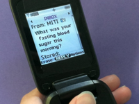
Background: Glucagon-like peptide-1 receptor agonists (GLP-1 RAs) and basal insulin both lower blood sugar, but while insulin puts people at risk of hypoglycemia and weight gain, GLP-1 RAs do not. In addition, GLP-1 RAs have added cardiometabolic and renal benefits. For these reasons, when possible, many primary care providers prefer their patients with type 2 diabetes to be from basal insulin to a GLP-1 RA. This transition process can be labor intensive, requiring multiple dosing adjustments and a watchful eye for hypoglycemia and hyperglycemia. The Mobile Insulin Titration Intervention (MITI)–GLP1 program uses SMS text messaging–based technology to support a streamlined and supervised transition process from basal insulin to a GLP-1 RA. This program takes place at a multilingual safety-net clinic. Objective: Our objectives were to assess program feasibility and acceptability to determine whether the intervention was doable, practical, and worthy of further investigation via a larger controlled trial. Preliminary clinical outcomes are also discussed in this paper. Methods: Patients were enrolled on a secure web platform that sent them a daily SMS text message asking the following: “What was your fasting blood sugar this morning?” Each weekday, texted responses containing patients’ fasting blood sugar levels were checked for alarm values, and once weekly, patients were called and advised on whether and how to lower their basal insulin and increase their GLP-1 RA dose. The program was co-run by general internal medicine physicians and nurses and continued until the patient had their insulin stopped completely and/or their GLP-1 RA dose reached the maximum, or 16 weeks elapsed. All enrolled patients were included in the analyses. Results: A total of 72 patients completed the pilot program. Feasibility and acceptability were high. Of 3671 SMS text messages sent by the program, 3520 (95.89%) received a response from patients. Of 719 cumulative weeks in which Thursday titration phone calls were attempted, successful connections with patients were made in 649 (90.26%) instances. Preliminary clinical outcomes were promising. Insulin doses were meaningfully reduced (55/72, 76.39% had their basal insulin reduced by at least 50%; 45/72, 62.5% had their insulin stopped completely). GLP-1 RA doses were meaningfully increased (64/72, 88.89% had their GLP-1 RA dose increased by ≥1 level; 45/72, 62.5% were discharged on the maximum dose of their GLP-1 RA). There was minimal hypoglycemia (5/3520, 0.14% of the SMS text messages reported a value of <80 mg/dL) and hyperglycemia (1/3520, 0.03% of the SMS text messages reported a value of >400 mg/dL). Conclusions: A general internal medicine–run MITI-GLP1 pilot program using SMS text messaging and interdisciplinary teamwork between internists and nurses is a feasible and acceptable intervention for safely and effectively transitioning people with well-controlled type 2 diabetes away from basal insulin and toward a GLP-1 RA.
A single infusion of zorpocabt agene-autoleucel (Zorpo-cel), an autologous CAR T-cell therapy, has led to a rapid and sustained remission in a patient with multiple life-threatening autoimmune disorders, according to a newly reported case published in Med.
Researchers from the University Hospital of Erlangen at Friedrich-Alexander-Universitat Erlangen-Nürnberg in Germany describe how a CD19-targeting CAR T-cell therapy successfully treated a 47-year-old woman suffering from severe autoimmune hemolytic anemia (AIHA), along with immune thrombocytopenia (ITP) and antiphospholipid antibody syndrome (APLAS). All three conditions are driven by malfunctioning B cells that produce harmful autoantibodies attacking the body’s own tissues.
The patient’s condition had proven exceptionally difficult to manage. Over nearly a decade, she had undergone nine different treatment regimens, including steroids, immunosuppressants, and antibody-based therapies, without lasting success. Her AIHA was particularly severe, leaving her dependent on daily blood transfusions and at risk of organ damage due to chronic anemia and iron overload.
With no effective options remaining, the clinicians, under compassionate use, turned to the CAR T-cell therapy developed by Miltenyi Biomedicine. This technique involves collecting a patient’s own T cells, genetically modifying them to target the B cell marker CD19 and reinfusing them to eliminate the dysfunctional immune cells.
The results were striking. Within just seven days of treatment, the patient no longer required blood transfusions. By day 25, her hemoglobin levels had returned to normal, indicating a complete resolution of the hemolytic anemia. Laboratory markers of red blood cell destruction also normalized rapidly.
Equally notable was the therapy’s broader impact. The patient’s elevated antiphospholipid antibodies—responsible for dangerous blood clots in APLAS—fell to normal levels and remained undetectable through 11 months of follow-up. Meanwhile, her platelet counts stabilized, indicating improvement in ITP without the need for additional treatment.
Researchers attribute this success to a “reset” of the patient’s B cell population. Unlike conventional therapies such as rituximab, which partially deplete B cells, CAR T cells appear to achieve deeper and more durable elimination. When B cells eventually returned months later, they were predominantly naïve, suggesting a reprogrammed and healthier immune profile.
Importantly, the treatment was well tolerated. The patient experienced none of the serious side effects commonly associated with CAR T therapy in cancer patients, such as cytokine release syndrome or neurotoxicity. Some mild liver enzyme elevations and blood count abnormalities were observed, likely related to prior treatments and iron overload rather than the therapy itself.
This is the second clinical win for Zorpo-cel in the treatment of autoimmune diseases this year. In January, the Phase I/II basket trial known as the CASTLE trial reported encouraging early results of Zorpo-cel administration in 24 patients with treatment-resistant autoimmune diseases, including systemic lupus erythematosus (SLE), systemic sclerosis (SSc), and idiopathic inflammatory myopathies (IIM). The therapy showed a favorable safety profile, with no cases of severe cytokine release syndrome or neurotoxicity observed. Efficacy outcomes were strong: 22 of 24 patients met predefined endpoints, including remission in most SLE patients, halted disease progression in all SSc patients, and meaningful clinical responses in the majority of IIM cases.
The case highlights the growing potential of CAR T therapy beyond oncology. Previous studies have shown promising results in systemic autoimmune diseases like lupus, but evidence in hematologic autoimmune disorders such as AIHA has been limited. While the findings are encouraging, researchers caution that this is a single case report. Larger, controlled clinical trials will be necessary to confirm safety, effectiveness, and long-term outcomes across diverse patient populations.
You’re reading the web edition of D.C. Diagnosis, STAT’s twice-weekly newsletter about the politics and policy of health and medicine. Sign up here to receive it in your inbox on Tuesdays and Thursdays.
In which RFK Jr. says the “government lies to us” while sitting in front of an HHS seal. Send news tips and podcast recommendations to John.Wilkerson@statnews.com or John_Wilkerson.07 on Signal.
First, control health care costs
Last month, a group of 12 Senate Democrats proposed a framework for rebuilding the health care system. The idea was to spur input from others ahead of when Democrats might get a chance to act on those plans.
Post-stroke insomnia (PSI) is a critical biological barrier to neurorehabilitation afflicting over half of all stroke survivors. Traditional sedatives often force clinicians into a therapeutic dilemma between sleep efficacy and cognitive suppression. The microbiota-gut-brain (MGB) axis has recently emerged as a transformative target to resolve this impasse. Acute stroke triggers profound autonomic dysfunction, causing immediate intestinal barrier collapse. This “leaky gut” facilitates the systemic translocation of lipopolysaccharides (LPS) and activates the NLRP3 inflammasome. The resulting inflammatory storm hijacks central tryptophan metabolism via the indoleamine 2,3-dioxygenase (IDO) enzyme. This “tryptophan steal” diverts serotonin precursors toward neurotoxic kynurenine pathways, driving severe cortical hyperarousal. Sleep fragmentation then prevents the glymphatic system from clearing metabolic waste, further exacerbating neuroinflammation. To break this vicious cycle of neurotoxicity, we propose a phase-dependent therapeutic framework. During the highly vulnerable acute phase, interventions must prioritize gut barrier protection using postbiotics to mitigate infection risks under CNS injury-induced immunodepression (CIDS), often discussed as stroke-induced immunosuppression. As patients enter the chronic phase, therapy shifts toward metabolic restoration using live therapeutics, such as washed microbiota transplantation (WMT) and next-generation psychobiotics like Akkermansia muciniphila. Targeting the MGB axis offers a mechanism-based strategy to achieve precision sleep medicine, restoring the biological foundation necessary for optimal neuroplasticity and recovery.
Get your daily dose of health and medicine every weekday with STAT’s free newsletter Morning Rounds. Sign up here.
A First Opinion essay today argues that “tastiness is not why people overeat.” But I may beg to differ — and so may a friend of mine who recently gave me a stuffed scolding after I convinced her to order pad thai for the table in addition to our meals.
On Thursday, the federal government closes the letter-of-intent window for MAHA ELEVATE, a $100 million initiative to fund “functional or lifestyle medicine” interventions for Medicare beneficiaries. The stated goal is to test evidence-based approaches to chronic disease prevention alongside conventional care. The unstated risk is that it will open a federal funding pipeline to interventions that sound integrative but can’t survive contact with a plausibility filter.
I’m a palliative care physician. I spend my days managing pain, breathlessness, nausea, and the existential weight of serious illness. My field should be cheering this investment in whole-person care. Instead, I’m watching it with one eye on the evidence and the other on a pattern I’ve seen up close: When uncertainty is high and emotions are higher, sectarian certainty moves in fast.
There is no doubt that autologous chimeric antigen receptor (CAR) T-cell therapy has revolutionized the treatment of serious blood cancers. A significant proportion of advanced-stage blood cancer patients who failed to respond to previous therapies now go into remission with this treatment, with some remaining cancer-free in the long term.
However, despite their success, these immunotherapies have significant disadvantages. Although CAR T-cell therapies have essentially rescued advanced-stage patients who previously would only have been offered palliative care, serious side effects such as cytokine release syndrome and immune effector cell-associated neurotoxicity syndrome (ICANS) are associated with the treatment.
All seven CAR T-cell therapies approved by the U.S. Food and Drug Administration (FDA) since August 2017 are autologous cell therapies, wherein the patient’s own T cells must be extracted and genetically engineered in the lab to produce cancer-targeting CAR T cells that are reinfused into the patient to fight the cancer.
Unless they live near a major cancer center or company with the relevant expertise and lab capacity in-house, many eligible patients miss out on the therapy because the wait time is too long or the whole process is too expensive. Patients must also be admitted to the hospital to undergo lymphodepletion chemotherapy before receiving the final infusion to allow the infused cells to expand, persist, and work better.
“It’s just simply too expensive, too complex to manufacture, and has all kinds of logistical issues that translate to limited patient access,” explained Maurits Geerlings, MD, co-founder, CEO, and president of in vivo CAR T-cell therapy biotech NanoCell Therapeutics, which has offices in Pennsylvania and Utrecht.
“Also, importantly, the batch capacity in highly specialized hospitals is so limited that altogether maybe 10% of patients that are eligible effectively get access to CAR T-cell therapy in the Western world.”
Maurits Geerlings, MD Co-founder, CEO NanoCell Therapeutics
Initially, after the first ex vivo, autologous CAR T-cell therapies like Novartis’s Kymriah and Kite’s Yescarta were approved in 2017, the field looked to develop “off-the-shelf” allogeneic therapies made from donor cells that would overcome some of the issues with autologous CAR T-cell therapies.
Despite the best efforts of a number of companies and researchers, no allogeneic CAR T-cell therapies have yet reached the market, although some companies like Allogene have reached Phase II trials. This is likely due to a few factors, such as adverse events linked to the rejection of donor cells, complex engineering problems, and the small margin of benefits of allogeneic over autologous CAR T-cell therapies.
Instead, over the last couple of years, the focus of the field has moved towards developing next-generation in vivo CAR T-cell therapies. Until recently, vectors or nanoparticles that could hit T cells precisely, safely, and predictably enough in humans to justify skipping ex vivo engineering were simply not available, but this is now changing.
In vivo CAR T-cell therapy uses the patient as a bioreactor. Upon injecting an engineered treatment carried by a vector such as a lentivirus or a lipid nanoparticle (LNP), it programs the patient’s T cells to attack either the cancer or autoreactive B cells in the case of autoimmune disease.
The field is still young, but initial clinical results reported last year in multiple myeloma blood cancer by Kelonia Therapeutics and in the B cell-driven autoimmune disease systemic lupus erythematosus by MagicRNA, as well as from EsoBiotec and academic labs, are promising.
Kevin Friedman, PhD Co-founder, CEO Kelonia Therapeutics
“It’s early days, so I don’t want to overinterpret the data. It’s also only in four patients, but what we are seeing is substantially better than what ex vivo CAR T cells have shown from an efficacy perspective,” emphasized Kelonia CEO and co-founder Kevin Friedman, PhD.
Indeed, this early success seems to have prompted intense investor and big pharma interest in the field. Since March 2025, when EsoBiotec was acquired by AstraZeneca, at least four other in vivo CAR T cell biotechs have been acquired, including Capstan Therapeutics by AbbVie and Interius BioTherapeutics by Kite/Gilead.
Whether in vivo CAR T-cell therapy will truly be the future of the field remains to be seen, but its convenience, economic viability, and the fact that it is effectively an “off-the-shelf” therapy that does not require lymphodepletion make it an attractive prospect for many.
Credit: Kelonia
Lentiviral vectors: Sticking with a known quantity
Five of the seven FDA-approved autologous CAR T-cell therapies, including Kymriah, use lentiviral vectors in the lab to engineer a patient’s T cells and transform them into CAR T cells.
Many of the most advanced companies in the in vivo CAR T- cell therapy space are applying similar technologies and using lentiviral vectors to target and transform T cells, but inside the body rather than in the lab.
Kelonia, which is based in Boston, is a leader in the in vivo CAR T-cell space and has already started clinical trials with its lead candidate KLN-1010 for the treatment of patients with relapsed and refractory multiple myeloma.
“It’s essentially delivering a fully human anti-BCMA (B cell maturation antigen) CAR to T cells, to reeducate them by expressing this anti-BCMA CAR inside the body to fight their tumor cells. Just like Abecma, or Carvykti, but it’s all done inside the body,” explained Friedman.
At the American Society of Hematology Annual Meeting in December last year, the company presented early Phase I results from four patients with relapsed and treatment-resistant myeloma who were treated with KLN-1010.
Although the study was small, the results were promising, with all four patients showing 100% minimal residual disease-negative response rate at follow-up and a lower rate of side effects than approved autologous CAR T-cell therapies.
“With our data and the efficacy and the safety profile that we’re seeing, this has a real shot at getting out of the major medical centers and into the community hospitals where the patients live, so they don’t have to travel to major medical centers,” said Friedman.
“Doctors can potentially treat patients in their own community and get access to the 90% of myeloma patients who, right now, despite the profound clinical benefit that CAR T cells provide, cannot be treated.”
Ryan Larson, PhD Senior Vice President Umoja Biopharma
Umoja Biopharma is another biotech using lentiviral vectors to develop in vivo CAR T-cell therapies. “We have three different products in the clinic currently. Two of those products are in B-cell malignancies, and one of the products is in autoimmune disease. And we’re making really great progress in enrolling patients across those studies,” said Ryan Larson, PhD, senior vice president and head of research at Umoja, although the Seattle-based company has not yet released any results from its Phase I studies.
Although Umoja is using lentiviral vectors, it has built in a rapamycin-activated cytokine receptor, which essentially acts as a booster switch for the engineered T cells in cancer patients while slightly dampening the rest of the immune system.
“It allows us to deliver a controlled pro-survival signal selectively to our CAR T cells in vivo,” explained Larson. “We’re able to potentiate persistence in a controlled manner in our in vivo generated CAR T cells with this rapamycin-activated cytokine receptor to drive persistence and ongoing immune surveillance, thus driving the key durable outcomes in oncology specifically.”
The requirements for autoimmune disease patients are different from those of advanced cancer patients, with a greater focus on safety. Long-term depletion of B cells is also not ideal, with the aim being to reset the immune system by getting rid of autoreactive B cells and replacing them with healthy ones.
“You’re eliminating all of the autoreactive repertoire and replacing it with a normal B cell repertoire, thus driving, ideally, a durable response wherein those autoimmune disease patients are no longer reliant on all the various immunosuppressants that are typically used to treat autoimmune disease,” said Larson.
Kite Pharma, now owned by Gilead and headquartered in California, was a pioneer in the autologous CAR T-cell therapy space. It developed Yescarta, one of the first two autologous CAR T-cell therapies approved by the FDA to treat blood cancers in 2017. Kite recently acquired Interius BioTherapeutics, a biotech in the lentiviral in vivo CAR T-cell space, for $350 million.
Priti Hegde, PhD Senior Vice President Kite Pharma, a Gilead Company
“The reason why we moved forward with Interius was that the clinical proof of concept for lentiviral-based delivery systems is far more advanced than for LNP-based systems,” said Priti Hegde, PhD, senior vice president and global head of research at Kite. “We were really excited to see that translation of the pharmacokinetics from an ex vivo platform to an in vivo platform.”
While lentiviral vectors are arguably “tried and tested” in the CAR T-cell space, there are some disadvantages associated with using them. For example, they can be hard to produce, implying that it is expensive and challenging to scale up manufacturing.
This is something both Umoja and Kelonia seem to have addressed, however. “We actually are quite unique from a biotech perspective in that we have our own, wholly owned manufacturing facility … It’s really allowed us to have a true pipeline from an in vivo cell therapy development perspective,” said Larson. “We’re actively working in our early phase clinical trials in a manufacturing setting that we know is scalable to commercial readiness.”
Kelonia does not do all its manufacturing in-house, but Friedman said that they have worked hard to develop a system that can be scaled. “Manufacturing is complicated. We like to think that we were thoughtful about our manufacturing approach, but it’s challenging generating these particles, these complicated medicines for Phase I use. We did it, though, and we now have a very reliable and scalable manufacturing process.”
Another potential risk linked to lentiviral and other viral vectors is that there is a small but significant risk of the vector inducing unwanted mutations in the DNA of target cells.
“Viral vectors have a propensity to integrate in transcriptionally active gene regions where you don’t want to go, because that enhances the mutagenesis risk,” noted NanoCell’s Geerlings.
Taking the non-viral route
Not everyone working to develop in vivo CAR T-cell therapies is using viral vectors. The second main route that companies and researchers are following to develop these cell and gene therapies is to use mRNA encapsulated in an LNP.
Last September, Shenzhen-based Chinese biotech MagicRNA published data from a Phase I trial of its in vivo mRNA and LNP-based CAR T-cell therapy in five patients with systemic lupus erythematosus. Similar to in Kelonia’s cancer trial, the results were promising. However, larger studies are needed for more conclusive results, as the sample size was small. But rapid, near-complete B cell depletion was seen for up to 10 days with no significant side effects like serious cytokine release syndrome or ICANS.
Since the pandemic, the use of mRNA therapeutics has become much more mainstream. For example, both of the prevalent vaccines against COVID-19 use a combined mRNA–LNP approach. In in vivo CAR T-cell therapy, the LNPs are used to take CAR-encoding mRNA to the right target cells in the body. Once inside a T cell, the LNP breaks apart and releases the mRNA into the cytoplasm. The cell’s protein synthesis machinery reads the mRNA and makes the correct CAR protein, which is then added to the surface of that T cell.
Aera Therapeutics, founded by CRISPR pioneer Feng Zhang, PhD, and based in Cambridge, Massachusetts, takes a combined mRNA–LNP approach to in vivo CAR T-cell therapy development, with a focus on treating B cell-mediated autoimmune disease.
Akin Akinc, PhD CEO, Aera Therapeutics
“We were really focused on autoimmune indications, so we said, ‘Let’s try to build a product profile that’s a great fit for that,’” explained Akin Akinc, PhD, who is CEO at Aera.
“You have the risk of insertional mutagenesis with lentiviral vectors. Even if those rates are small, they’re not zero … So that’s why we thought an mRNA–LNP approach, where there’s no chance of insertion, is theoretically a more attractive approach.”
Aera has not yet moved into clinical studies but reported preclinical data for its therapy candidate AERA-109 in non-human primates at the American Society of Hematology Annual Meeting at the end of last year. They showed potent and durable B cell depletion across different tissues in the body.
One potential disadvantage of using a combined mRNA–LNP approach, particularly for treating cancer, is that it is unlikely to last as long as a lentiviral approach. As this could be potentially advantageous in people with autoimmune disease, where B cell depletion does not need to occur over such a long period of time, it seems to be the most common method followed by companies designing in vivo CAR T-cell therapies for autoimmune conditions.
“Our therapeutic goal is to go in and clear out the B cells that exist in the body, both in the periphery and the tissues, and then allow them to repopulate. If we achieve that immune reset, then that’s all that we can do,” said Akinc.
Scott Barros, Head of Early Development, Akin Akinc, Chief Executive Officer, Bill Querbes, Chief Scientific Officer.
“Then the question is, is there going to be a relapse 12–18 months later? But so long as we clear out all the B cells, which happens pretty quickly, I think there’s no benefit to having the CAR T cells hanging around for longer, because at that point you’ve done the job. Then it’s about whether or not that remission is going to persist.”
NanoCell Therapeutics is also taking a non-viral approach to developing in vivo CAR T-cell therapy for treating B-cell malignancies, but is using DNA instead of RNA. The candidate has not yet reached the clinic, but it has achieved good preclinical results and will soon be tested in non-human primates.
Similar to Aera, NanoCell packages its therapy in targeted LNPs. However, these carry a minicircle DNA that encodes the CAR information and an mRNA transposase that allows the DNA to integrate into the target cell genome.
“We still remain, I think, pretty much in the lead as a company delivering non-viral DNA, because it is very difficult … We see an opportunity for us to actually make a breakthrough there,” said Geerlings.
“The nuclear membrane of the cell is such a barrier. You need to find an opportunity to open it up and to be just in time with your DNA in a way that is not triggering an innate immune response. You also need to have a mechanism by which that DNA can integrate, because otherwise, you will end up having an episomal expression of your DNA.”
The approach taken by NanoCell is definitely at an earlier stage than the lentiviral and mRNA–LNP approaches that are already generating clinical data, but there are a couple of other companies working on similar products, like Stylus Medicine and CPTx. If it works, then this approach has the promise of ruling out problems with viral vectors, such as manufacturing difficulties. It would also theoretically generate longer-lasting and more durable treatment effects than could be achieved with mRNA.
What’s next for CAR T-cell therapy?
It seems that we are on the cusp of next-generation in vivo CAR T-cell therapies, although the studies published so far have all been small and it remains to be seen if the current buzz in the space is based on hype or reality.
“I do think that in vivo will go from a platform with initial proof-of-concept to broad applicability faster than perhaps ex vivo platforms did,” said Hegde. “But we have a lot of scientific questions. For example, in the absence of lymphodepletion, can an in vivo platform give you the depth and durability of response that an ex vivo platform does?”
There is a lot of interest in whether safer and more accessible in vivo CAR T-cell therapy can make this treatment approach more appealing to people with B-cell-mediated autoimmune conditions than autologous CAR T-cell approaches. The initial clinical results are good, but questions remain about how long the results will last.
“I think these are going to work, but we’re going to learn things that allow us to make second-generation products that are even better and even more potent,” said Akinc.
On the cancer side of things, most companies developing in vivo CAR T-cell therapies for oncology indications are sticking with blood cancers against which autologous CAR T-cell therapies have already been shown to be efficacious.
“I think we’ll continue to see strong proof of concept in de-risked indications like the hematologic malignancies over the next year,” said Larson. “Over the next two years, I think we’re going to be closely watching the field for durable outcomes in oncology and the ability to drive immune reset in autoimmune disease that then translates to durable remissions in autoimmune disease patients.”
A big question on everyone’s mind is whether this new technology could help overcome some of the hurdles that prevent CAR T-cell therapy from being successful at treating “solid” tumors, such as working out how to overcome diverse tumor microenvironments.
“I think there’s great potential in solid tumors. One reason why we went with Interius was [that] we think that the application of the in vivo platforms could really break open the problems that we perhaps had in solid tumors with ex vivo CAR T cells,” said Hegde.
“The nice thing about the in vivo space is you can put whatever targeting antigen you want on the virus to go to a specific cell type. So it’s really up to your imagination, how you want to design an in vivo CAR T cell.”
Dispatch Bio is also in the CAR T-cell therapy space, but is targeting solid tumors rather than developing in vivo CAR T cells. The company is based in Philadelphia and was co-founded by Carl June, MD, one of the pioneers of CAR T-cell therapy.
The technology they are developing is a two-component system, in which a human-specific adenovirus designed to infect cancer cells, but not healthy tissue, is used to “paint” the tumor cells so that a CAR T cell can more easily home in on the cancer and destroy it.
Barbra Sasu, PhD Chief Scientific Officer Dispatch Bio
“The virus gets into the tumor microenvironment and then, because it’s a virus, creates an inflammatory condition. When it does that, it’s immediately more supportive for T cells,” explained Dispatch chief scientific officer Barbra Sasu, PhD.
“We’re doing what T cells can’t do for themselves. We’re expressing a target and directing them to kill what we want them to kill. We’re also adding a cytokine to support them and, actually, the endogenous immune system too.”
The two-part approach is very new, so Sasu and colleagues are testing the system using a known autologous CAR T-cell therapy approach. But she says that the system is potentially very flexible and could allow a wide range of therapies, including in vivo CAR T-cell therapies, to be combined with the viral targeting approach if they prove effective.
“What we wanted to do was to start with something that we felt we understood very well. We also had the benefit in our first program of being able to work with people who’ve already developed CAR T-cell therapies,” said Sasu. “That’s a big advantage because [when] coming in with a two-component system, it’s good if you don’t have to refine both parts at once.”
Another CAR T-based approach being developed by Kite and others in this space is logic gating, which is the development of IF, AND, and NOT switches to allow much more refined control of CAR T-cell therapies by clinicians and potentially increase effectiveness in complex solid tumors.
“We’re really interested in exploring the logic gating space and what it can do to deliver CAR T cells more safely, especially in solid tumors, where the antigens aren’t as broadly homogeneously expressed,” said Hegde.
Helen Albert is senior editor at Inside Precision Medicine and a freelance science journalist. Prior to going freelance, she was editor-in-chief at Labiotech, an English-language, digital publication based in Berlin focusing on the European biotech industry. Before moving to Germany, she worked at a range of different science and health-focused publications in London. She was editor of The Biochemist magazine and blog, but also worked as a senior reporter at Springer Nature’s medwireNews for a number of years, as well as freelancing for various international publications. She has written for New Scientist, Chemistry World, Biodesigned, The BMJ, Forbes, Science Business, Cosmos magazine, and GEN. Helen has academic degrees in genetics and anthropology, and also spent some time early in her career working at the Sanger Institute in Cambridge before deciding to move into journalism.
Neuroblastoma is the most common tumor among children under a year of age, and while in its gentlest form neuroblastoma can regress on its own, it can also take an aggressive form, with high-risk neuroblastoma carrying a five-year survival rate of about 40%.
Researchers at The Hebrew University of Jerusalem have now discovered a mechanistic explanation for how neuroblastoma sustains itself and identified a potential approach to severing that mechanism, by inhibiting nitric oxide (NO) production to suppress mTOR signaling. The collective results from work in human neuroblastoma cells and experiments in a mouse xenograft model showed that inhibiting the enzyme neuronal nitric oxide synthase (nNOS) to inhibit NO production suppressed mTOR signaling and slowed tumor growth.
Professor Haitham Amal, PhD, head of The Laboratory of Neuromics, Cell Signaling, and Translational Medicine, is senior and co-corresponding author of the team’s published paper in Brain Medicine, titled “Targeting nNOS suppresses AKT–TSC–mTOR signaling and inhibits neuroblastoma growth.” In their paper the team concluded “Inhibition of nNOS suppresses mTOR signaling, reduces cellular malignancy, and attenuates tumor growth in vivo, identifying the nNOS-mTOR axis as a promising therapeutic target in neuroblastoma.”
Neuroblastoma accounts for roughly 28% of all cancers diagnosed in infants across Europe and the United States. “Neuroblastoma (NB) refers to a spectrum of neuroblastic tumors that originate from the neural crest cells during fetal development,” the authors wrote. “Neuroblastoma is predominantly a pediatric malignancy, with approximately 97% of cases occurring in children.”
NBs can range from spontaneous regression to maturation to an aggressive, deadly metastatic disease. And as the investigators noted, “Despite major advances in multimodal therapy, high-risk neuroblastoma remains associated with poor prognosis, frequent relapse, and therapy resistance, underscoring the need for a better understanding of the signaling pathways that regulate tumor cell survival, differentiation, and metabolic adaptation.”
Nitric oxide (NO) is an essential regulator of carcinogenesis in various tumors, including NB, the authors pointed out. “Nitric oxide (NO) is a ubiquitous free radical signaling molecule produced in multiple organs and tissues), such as those of the central and peripheral nervous systems.” But at elevated concentrations NO becomes reactive, generating nitrogen species that chemically modify proteins through a process called S-nitrosylation. That modification has been implicated in every stage of cancer progression.
The relationship between nitric oxide and tumors is not simple. Very high concentrations can damage DNA and trigger apoptosis. Lower, sustained levels appear to do the opposite, promoting survival and metastasis. Amal and colleagues had previously demonstrated that nitric oxide drives glioblastoma progression. The question that remained was whether the same enzyme, neuronal nitric oxide synthase, was performing a similar service for neuroblastoma, and if so, through which downstream pathway. The answer turned out to be mTOR.
The team attacked nNOS from two directions. They treated human SH-SY5Y neuroblastoma cells with BA-101, a selective pharmacological inhibitor, at 100 μM for 24 hours. Separately, they silenced the nNOS gene with small interfering RNA. The reasoning was that if a drug and a genetic tool produce the same result, you are looking at biology, not pharmacological noise.
The experiments produced the same result. BA-101 reduced NADPH-diaphorase activity, the standard readout of NOS function, by 35-40%. Genetic silencing cut it by 45-50%. Nitrite levels, a stable proxy for nitric oxide production, fell 65-70% with BA-101 and 55-60% with siRNA. Colony formation, the most direct measure of proliferative capacity, dropped significantly after both BA-101 treatment (p < 0.001) and nNOS silencing (p < 0.01). The cells were losing their ability to multiply.
What followed downstream was systematic. Protein tyrosine nitration, measured by 3-nitrotyrosine immunoreactivity, fell sharply after BA-101 treatment (p < 0.01) and nNOS silencing (p < 0.001). The chemical signature of nitrosative stress was fading.
The results then confirmed that AKT phosphorylation decreased (p < 0.01 with BA-101; p < 0.05 with siRNA), while total AKT remained unchanged. Phosphorylation of mTOR itself declined under both conditions (p < 0.01 each). The downstream mTORC1 substrate ribosomal protein S6 followed (p < 0.05 with BA-101; p < 0.01 with siRNA).
And here, the most telling detail, that TSC2, a master negative regulator of mTOR signaling, rose significantly under both treatments (p < 0.05). Removing the nitric oxide signal had allowed the cell’s own braking system to re-engage. In summary, the authors noted, “Pharmacological inhibition of nNOS with BA-101 (100 μM, 24 h) or genetic silencing of nNOS with siRNA caused upregulation of the key negative regulator TSC2 and decreased phosphorylation of AKT, mTOR, and RPS6, indicating suppression of mTOR pathway activity.”
Synaptophysin, a neuroendocrine tumor marker used to gauge the malignant identity of neuroblastoma cells, decreased significantly with BA-101 (p < 0.01) and nNOS knockdown (p < 0.05). The tumor cells were not merely growing more slowly. They were becoming, at a molecular level, less recognizably cancerous. In summary, the investigators noted, “Our results show that inhibition of NO production in the human NB cell line (SH-SY5Y cells), either by pharmacological intervention using the selective nNOS inhibitor BA-101 (41) or by genetic ablation using the specific siRNA, successfully suppressed NB malignancy.”
Schematic model illustrating the NO-mTOR signaling axis in neuroblastoma. Under basal/pathological conditions (left panel), and nNOS inhibition (right panel). [Haitham Amal]
But if blocking nitric oxide suppresses mTOR signaling, then flooding the cell with nitric oxide should amplify it. The researchers tested this by exposing SH-SY5Y cells to SNAP, a nitric oxide donor, at 200 μM for 24 hours. This converse experiment produced the converse result. 3-nitrotyrosine rose (p < 0.05), and TSC2 fell (p < 0.01). Phosphorylation of AKT, mTOR, and RPS6 all increased (p < 0.05 for each).
The team then tested their findings in a xenograft mouse model of neuroblastoma, treated with BA-101. “Importantly, to extend these findings to an in vivo context, we further assessed the impact of pharmacological nNOS inhibition on tumor growth in a xenograft NB model,” they stated. The investigators found that while tumors in control animals grew to approximately 1.5 cm in their largest dimension, the treated tumors did not. Final tumor volume and weight were dramatically reduced in the BA-101 group. ‘Quantitative analysis revealed a dramatic decrease in the final tumor volume and weight in the BA-101-treated group (p < 0.001) compared with controls,” they noted.
Body weight did not differ significantly between groups, suggesting that the compound was tolerated without gross systemic toxicity. In summary, the authors wrote, “Our finding demonstrate that the pro-tumorigenic effects of nNOS in SH-SY5Y involve activation of themTOR signaling pathway.” Importantly, both genetic inhibition of nNOS using siRNA and pharmacological inhibition with BA-101 effectively suppressed mTOR pathway activation and reduced malignant properties of NB cells, highlighting the therapeutic relevance of targeting nNOS signaling. “These findings indicate that pharmacological inhibition of nNOS effectively suppresses xenograft tumor progression, highlighting the critical role of nNOS-derived NO in promoting neuroblastoma growth in vivo.”
“The magnitude of the in vivo suppression caught our attention,” said Amal, the study’s corresponding author, who holds appointments at the Institute for Drug Research, School of Pharmacy, Faculty of Medicine, The Hebrew University of Jerusalem, and the Rosamund Stone Zander and Hansjoerg Wyss Translational Neuroscience Center at Boston Children’s Hospital, Harvard Medical School. “We had demonstrated the role of nitric oxide in glioblastoma previously, but the consistency of the neuroblastoma results across every assay, from protein phosphorylation to colony formation to xenograft growth, points to nNOS as something more than a contributor. It appears to be a central driver of the signaling that sustains this tumor.”
Added first author Shashank Kumar Ojha, PhD, first author of the study and a researcher at the Institute for Drug Research, The Hebrew University of Jerusalem, added, “What convinced me was the concordance between the pharmacological and genetic approaches. When BA-101 and siRNA independently produce the same pattern of effects across NADPH-diaphorase activity, nitrosative stress markers, mTOR pathway phosphorylation, and clonogenic growth, you can be confident the biology is real. That reproducibility is what gives you a therapeutic hypothesis worth testing further.”
The authors acknowledged limitations to their study. The in vitro work relied on a single cell line, SH-SY5Y, which cannot capture the full genetic heterogeneity of neuroblastoma or the complexity of the tumor microenvironment. The chemical identity of BA-101 is currently undisclosed pending patent issuance, which means independent replication by other laboratories must wait. Whether nitrosative stress directly underlies its functional impairment, or whether an intermediary mechanism is involved, remains an open question that the authors explicitly flag for future investigation. “Future studies using patient-derived cells, organoids, or genetically engineered mouse models will be important to further validate and extend these observations,” they stated. Nevertheless, the authors suggest, the limitations do not diminish the central discovery of a druggable nNOS–mTOR axis.
mTOR inhibitors such as rapalogs and catalytic mTOR inhibitors have shown limited efficacy as monotherapies in neuroblastoma, undermined by feedback activation and resistance mechanisms. The present study suggests the potential for a different attack strategy. Rather than targeting mTOR at the lock, intervene upstream at the hand that turns the key. By reducing nitric oxide-dependent mTOR activation, nNOS inhibition may sidestep the compensatory pathways that have frustrated direct mTOR blockade. “Collectively, these results identify the nNOS-mTOR axis as a key driver of neuroblastoma progression and suggest that nNOS inhibition represents a promising strategy for NB treatment,” they concluded.




![Schematic model illustrating the NO-mTOR signaling axis in neuroblastoma. Under basal/pathological conditions (left panel), and nNOS inhibition (right panel). [Haitham Amal]](https://www.genengnews.com/wp-content/uploads/2026/04/Low-Res_Amal-Figure1-2026-Screenshot-2026-04-01-at-11.41.51-300x153.jpg)