Women who undergo medically assisted reproduction (MAR) may have a slightly higher risk of developing certain hormone-related cancers, but a large Australian study suggests much of that increase is likely explained by underlying infertility, greater medical surveillance, and other patient characteristics rather than the fertility treatments themselves.
The findings, published in JAMA Network Open, analyzed data from nearly 1.75 million Australian women and represent one of the largest investigations to date of cancer risk following fertility treatment. Using an emulated target trial design—a statistical approach intended to better approximate the conditions of a randomized clinical trial—the researchers examined whether three common forms of MAR were associated with later cancer diagnoses.
The study included 1,748,927 women aged 18 to 55 years between 1991 and 2018, including 396,661 who had received some form of MAR. Treatments evaluated included assisted reproductive technology (ART), intrauterine insemination (IUI) or ovarian stimulation, and ovulation induction with clomiphene citrate.
Researchers found modest increases in the relative risk of several hormone-related cancers—including breast, ovarian, uterine, thyroid, colorectal cancers, and melanoma—after some fertility treatments. However, the absolute increase in risk was small.
For any individual invasive cancer, the investigators estimated that treated women experienced fewer than 20 additional cancers per 100,000 women per year compared with women who had not undergone MAR.
The authors emphasized that the observed associations should not be interpreted as evidence that fertility treatment causes cancer. “Although we observed increased relative risk for most hormone-related cancers following MAR, this corresponded to only small increases in estimated absolute excess risk,” the authors wrote.
To better distinguish treatment effects from other influences, the investigators incorporated several bias analyses rarely included in previous studies. They calculated E-values to estimate the impact of unmeasured confounding and analyzed cancers not believed to be hormonally driven—including pancreatic, lung, and hematologic cancers—as negative controls.
Those analyses suggested that underlying infertility-related conditions, such as endometriosis and polycystic ovary syndrome, as well as factors including obesity, anovulation, and demographic differences, could account for much of the increased risk observed for ovarian, uterine, and thyroid cancers.
The researchers also found that cancer diagnoses tended to cluster during the first few years after fertility treatment. According to the investigators, “Several emulated trials suggested a greater likelihood of incident cancer shortly after first treatment.” They noted that this pattern could reflect either accelerated growth of preexisting cancers or increased medical monitoring during fertility treatment and pregnancy.
After examining the data, the authors concluded that enhanced surveillance was the more likely explanation. “We believe detection bias is the more likely explanation for these results. This finding is the first empirical indication using a negative control that medical surveillance may be responsible for an increased risk of cancer following MAR,” they wrote.
Women pursuing fertility treatment often undergo repeated medical evaluations and may also be more likely to participate in cancer screening programs, increasing the likelihood that existing cancers are detected earlier than they otherwise would be.
Overall, the researchers concluded that while associations between MAR and hormone-related cancers were observed, the evidence does not support a simple causal relationship.
The investigators said the findings should reassure patients while also encouraging careful counseling and follow-up. They recommend that clinicians discuss the possibility of a small increase in cancer risk but explain that the excess risk “may be partially or fully due to the health and sociodemographic profile of women who receive MAR and increased surveillance during treatment.”
The authors added that routine surveillance after fertility treatment remains appropriate, but they cautioned that observed increases in cancer diagnoses should be interpreted within the broader context of infertility-related health conditions and differences in healthcare utilization rather than being attributed solely to fertility medications themselves.
Nebius, an AI cloud company, sponsored its second “AI Discovery Awards” event and dinner earlier this month in London, where the winning companies were announced. The event highlighted leading startups in biopharma, genomics, medical devices, and digital health that are using AI to deliver advances in healthcare and life sciences.
During the evening awards ceremony, artificial intelligence demonstrated that it is rapidly reshaping biomedical research. However, practitioners agree that AI’s success depends on more than advanced algorithms. [Nebius]
At a Nebius-hosted morning discussion before the awards dinner, several researchers highlighted the importance of powerful computing infrastructure, high-quality biological data, and laboratory validation.
Examples included AI models that predict osteoarthritis years before symptoms and an Alzheimer’s platform, which achieved 97% diagnostic accuracy when paired with protein biomarkers. A Stanford Medicine scientist described CRISPR-GPT, an AI assistant that helps design and troubleshoot gene editing.
The investigators also spotlighted AI-powered lab automation, multimodal datasets, and AlphaFold’s dramatic acceleration of protein structure prediction. Across every application, participants emphasized that collaboration among academia, healthcare, industry, and governments will be essential to advance preventive, personalized medicine and to translate AI discoveries into clinical practice.
Ilya Burkov, PhD, who has a background in clinical medicine, is now global head of healthcare and life science at Nebius. Burkov began his research career focusing on osteoarthritis, osteoporosis, hip and knee replacements, and trying to figure out how such diseases develop and progress.
“My goal was to work backward from the end stage of disease and determine whether we could predict who was at risk years before serious joint damage occurred,” he explained. “If we could identify those patients early enough, perhaps we could delay disease progression.”
Machine learning
Speaking with colleagues in a hospital, he was asked: “Have you looked at it from any machine learning perspective?” Burkov had no formal background in artificial intelligence, but he was intrigued by the idea of using emerging AI models and advanced algorithms to analyze long-term medical imaging data.
Ilya Burkov, PhD [Nebius]
The concept was simple but powerful: if AI could identify patterns shared by patients who later developed osteoarthritis or osteoporosis, it might be able to detect subtle biomarkers long before the disease became clinically apparent.
“That idea became the focus of my PhD research. AI models were not a thing ten years ago when I was in the hospital. There were transformer models and algorithmic-based approaches.
“I developed techniques capable of predicting the early onset of osteoarthritis and osteoporosis with an accuracy of roughly 80% to 90%,” he pointed out. “The models identified imaging features that consistently appeared years before patients required joint replacement surgery.
“This made it possible to examine scans from otherwise healthy individuals and estimate their future risk. In some cases, we could tell patients that, without changes to certain lifestyle factors, they had a high probability of requiring a hip replacement within the next 10 to 15 years.”
For Burkov, that was transformative. AI made it possible to move beyond treating individual patients and instead create tools that could benefit entire healthcare systems. Rather than applying clinical expertise one patient at a time, scalable technologies could be created capable of helping clinicians identify high-risk patients earlier and intervening before irreversible damage occurred.
That realization ultimately convinced him to transition from clinical medicine into industry, where he saw the opportunity to build technologies that could have a much broader impact. Whether it’s a small academic lab with only a handful of researchers or a global pharmaceutical company operating at massive scale, every organization faces different computational challenges.
“At Nebius, our role is to provide the computing infrastructure and technology that enables researchers to train increasingly sophisticated AI models and accelerate scientific discovery to advance biomedical research and improve patient care,” he said.
Alzheimer’s disease
Artificial intelligence is rapidly reshaping drug discovery, but many researchers believe the greatest challenge is not designing drugs—it’s knowing what biological targets to pursue.
Prima Mente, a previous AI Discovery Award winner, is tackling that problem by building foundation AI models designed to uncover the molecular mechanisms behind Alzheimer’s disease and other neurodegenerative disorders. By combining blood-based biomarkers, multimodal biological data, and transformer-based AI, the London startup hopes to identify the molecular drivers of neurodegenerative disease—and ultimately accelerate the development of new therapies.
“If we can diagnose disease earlier, better stratify patients, and understand what’s actually driving Alzheimer’s, we can help create better treatments,” said co-founder Hannah Madan, PhD.
Based in London’s King’s Cross innovation district, Prima Mente has grown to approximately 35 employees across London, San Francisco, and the United Arab Emirates. Madan, whose academic background includes a master’s degree in pharmacology and a PhD investigating the relationship between bowel cancer and diabetes, has spent most of her career building biotechnology startups. Prima Mente is the fifth company she has helped launch.
The company’s mission addresses one of healthcare’s most pressing challenges. Dementia is the leading cause of death in the U.K. and the sixth highest in the U.S. Alzheimer’s disease remains the most common form of dementia worldwide.
Looking beyond traditional biomarkers
Prima Mente’s scientific strategy draws inspiration from advances in cancer diagnostics, particularly liquid biopsy technologies that detect circulating tumor DNA in blood samples. The company wondered whether a similar approach could work for neurodegenerative disease.
“When we started three years ago, many people thought we were a little crazy,” noted Madan. “The prevailing view was that very little DNA from dying brain cells entered the bloodstream.”
Hannah Madan, PhD [Nebius]
The team has since demonstrated that cell-free DNA originating from neurons, microglia, and astrocytes can be detected in blood. More importantly, those DNA fragments retain epigenetic information that may reveal the biological state of brain cells before they died.
Rather than focusing solely on DNA sequences, Prima Mente analyzes methylation patterns carried on cell-free DNA. Because methylation reflects how genes are regulated within specific cell types, these signals can provide insight into disease progression and cellular dysfunction.
“When cells die, they release fragmented DNA into the bloodstream,” Madan explained. “Those fragments preserve methylation signatures that tell us what state those brain cells were in.”
The biological strategy is paired with an equally ambitious computational one. Prima Mente believes that transformer architectures—the same AI technology underlying large language models such as ChatGPT—can learn the language of biology.
“If ChatGPT can understand human language, our hypothesis is that similar models can understand biological languages,” noted Madan.
Instead of converting sequencing data into simplified numerical counts, the company trains models directly on raw biological sequences, including DNA, methylation signals, RNA transcripts, and proteomic data. By preserving more of the underlying biological information, Prima Mente believes its models could uncover relationships that conventional bioinformatics pipelines often overlook.
The company’s first foundation model, known as Pleiades 1, demonstrated the potential of that approach. Initially trained to identify Alzheimer’s disease from blood-derived molecular data, the model successfully diagnosed a subset of patients. After protein biomarkers were incorporated, diagnostic accuracy increased to approximately 97% within the study dataset—exceeding the performance of current clinical standards, according to Madan.
AI tokens are the fundamental units of data processed by AI models during training and inference. They represent smaller components of text, audio, images, or other modalities, enabling models to understand, predict, and generate outputs effectively. Pleiades 1 was trained on 1.9 trillion tokens. Its successor, Pleiades 2, is being trained on 80 trillion tokens spanning five biological data modalities, with the long-term goal of building a 100-billion-parameter foundation model.
Prima Mente partnered with AI infrastructure provider Nebius, which supplied a dedicated 32-node computing cluster powered by NVIDIA GPUs. The additional computing capacity enabled the company to scale from a 1-billion-parameter model to a 10-billion-parameter model within weeks while increasing training throughput from roughly 8,000 tokens per second per device to more than 1.1 million tokens per second across a 16-node cluster.
Beyond model development, Prima Mente is collaborating with the U.K.’s National Health Service (NHS) through the Sandbox Study, which collects blood samples from patients with suspected neurological disease. The real-world data help researchers develop AI models aimed at detecting Alzheimer’s earlier, potentially enabling treatment before irreversible brain damage occurs.
Dementia is the leading cause of death in the U.K. and the sixth highest in the U.S. Alzheimer’s disease remains the most common form of dementia worldwide. [Cecille Arcurs/Getty Images]
Unlike AI companies that rely primarily on public datasets, Madan said Prima Mente is generating much of its own training data. The company collaborates with 20 NHS memory clinics throughout the U.K., collecting blood samples, speech recordings, clinical notes, and imaging data from patients at the earliest stages of cognitive decline. It also participates in the U.K.’s Sovereign AI initiative.
Lab validation is integrated into the company’s development process. Candidate discoveries generated by AI models are tested using stem cell systems, brain tissue, and additional blood-based experiments, creating a continuous feedback loop between computational prediction and experimental validation.
That combination of proprietary data generation, lab experimentation, and AI model development represents what the company views as a significant competitive advantage.
While AI has attracted enormous attention for accelerating drug discovery, Madan argues that identifying the right biological target remains the industry’s greatest bottleneck. She compares today’s AI revolution to the impact AlphaFold had on protein structure prediction. As computational tools become increasingly capable, designing drug candidates may become faster, cheaper, and more routine.
“But if you don’t know what biology actually matters,” she said, “there’s little value in having better tools to build drugs.”
For Prima Mente, Madan says the opportunity lies upstream of drug development—discovering the cellular pathways, biomarkers, and molecular mechanisms that should become tomorrow’s therapeutic targets.
That strategy recently received external validation when the company won the AI Insights Prize for Alzheimer’s from the Gates Foundation, receiving $1 million to expand research into microglial biology. The funding will support AI models designed to identify gene perturbations in specific brain cell types that could serve as the basis for future Alzheimer’s therapies.
As foundation models continue to expand beyond language into biology, Madan is betting that the next major AI breakthrough in medicine will not simply generate better drugs—it will reveal entirely new biology that makes those drugs possible.
CRISPR-GPT
CRISPR-GPT is a large language model developed by Stanford Medicine to automate key steps in CRISPR gene-editing research. Acting as an AI agent, it interprets scientific literature, designs guide RNAs, suggests experimental parameters, and integrates with lab automation systems to execute and refine experiments. By reducing manual planning and accelerating iterative testing, the system enables researchers to complete complex gene-editing workflows more efficiently and consistently, noted Stanford researchers.
CRISPR-GPT is also credited with lowering the barrier for scientists with limited CRISPR expertise, improving accessibility. The platform represents an emerging class of AI tools that can “reason” through complex scientific tasks, recommend next steps, and accelerate discovery. Potential applications include developing gene therapies, improving cancer research, engineering cell therapies, and expanding access to genome-editing technologies.
The goal, according to Le Cong, PhD, assistant professor of pathology and genetics is to help scientists produce life-saving drugs faster. “The hope is that CRISPR-GPT will help us develop new drugs in months instead of years,” he said.
Le Cong, PhD [Stanford Medicine]
Cong and team developed CRISPR-GPT using Nebius AI Cloud as its core infrastructure. The group leveraged Nebius’ GPU clusters to train their specialized CRISPR-Llama3 model, rapidly iterate on architectures, and scale from prototyping to full model training.
CHAT-GPT could also expand the pool of scientists who can effectively use gene editing technology—no experience required, pointed out Cong. “Trial and error is often the central theme of training in science, but what if it could just be trial and done?” he added. Cong is the senior author of a study “CRISPR-GPT for agentic automation of gene-editing experiments,” published July 2025, in Nature Biomedical Engineering.
AI Discovery Awards
At the AI Discovery Awards dinner in the evening, the sponsors announced that the 2026 program added medical devices and medical imaging to the existing biopharma, genomics, and digital health tracks to reflect the growing role of AI in connected medical equipment and diagnostic imaging.
“Our winners—and indeed all of the 647 submissions we reviewed—reflect how rapidly AI is changing the pace of healthcare research,” said Ilya Burkov during a short presentation. “Across all categories, startups are compressing timelines that once took years into months or even weeks, and bringing capabilities to clinical and laboratory settings that simply did not exist before.
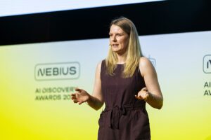
Margaret Hua, founding chief of staff at Phylo, accepts $100,000 in GPU cloud credits for first prize in the biopharma category. The company is building AI research assistants that can independently help biomedical scientists think through problems, design experiments, analyze data, and suggest what to do next, with the aim of speeding up scientific and biomedical discovery. [Nebius]
“The AI Discovery Awards exist to accelerate that momentum, and to connect the most promising teams with the compute resources, investor networks, and mentorship they need to move from promising research to bringing products to market.”
Alongside the awards program, Nebius previewed its Nebius Scientific AI and Healthcare Platform, which is an AI infrastructure built to meet the specialist needs of healthcare and life sciences organizations, explained a Nebius official.
The 2026 AI Discovery Awards were open to companies from pre-seed through to Series D that put AI and machine learning at the core of their product. Category winners were selected from 647 applications from around the world by an independent panel of 28 judges representing leading pharmaceutical companies, academic institutions, and venture capital firms. Submissions were evaluated based on the use of AI within the product, use of compute, technical innovation, functionality and advantages, performance and efficiency, global impact, market potential, and business sustainability.
A full list of shortlisted companies, as well as qualification criteria and a jury list, can be found on Nebius’s website.
Wearable electronic “tattoos” painted onto the skin could allow comfortable and reliable health monitoring to personalized designs.
The flexible electrodes, outlined in PNAS, represent a pathway to the next generation of wearable systems for healthcare.
The novel devices are made of intrinsically conductible polymers that conform to the skin and maintain reliable connections to electronic systems, allowing stable electrophysiological monitoring.
Their ability to create bespoke designs could be particularly useful for long-term use in children, adolescents, and people sensitive to potential stigmatization.
WE-PDD electrode [Wanqing Zhang]
The researchers describe their invention “as a personalized and scalable solution for next-generation electronic tattoos that combine ultra conformality, customizability, and long-term reliability, accelerating their translation into clinical diagnostics and interactive bioelectronic systems.”
The current gold-standard electrodes of wet silver/silver chloride gels can dry out and produce inaccurate readings during motion.
The team therefore developed a paintable, drawn-on-skin dry electrode based on the biocompatible polymer poly (3,4-ethylenedioxythiophene): poly (styrene sulfonate (PEDOT:PSS).
The conductive ink can be painted onto microtextured skin with a commercial paintbrush and even go through hair.
It comprises polyvinyl alcohol as a mechanically supportive network, PEDOT:PSS as a conductive filler, and 4-dodecylbenzenesulfonic acid (DBSA) as both conductive additive and plasticizer, in a water-ethanol cosolvent system.
The resulting WE-PPD (water-ethanol-PVA/PEDOT:PSS/DBSA) electrode is breathable, sticks well to the skin, and conforms to it closely, minimizing skin contact impedance.
The paintable formulation also enables it to penetrate porous silver textile to form a robust soft-rigid connection.
In proof-of-concept demonstrations, the team demonstrated the versatility of the electrodes for biopotential monitoring and human-machine interfaces.
It was tested across wireless long-term electrocardiography recordings, robotic hand control based on electromyogram, and electroencephalogram-based neural response detection.
The absence of imaging artifacts also demonstrated its potential for multimodal MRI imaging and electrophysiological (EP) recordings, noted first author Wanqing Zhang, a PhD research assistant at Penn State University, and co-workers.
The team added: “The ability to customize the electrode appearance transforms WE-PPD from a conventional medical device into a user-friendly and aesthetically integrated wearable technology.
“For example, electrodes can be designed with cartoon patterns, which may reduce anxiety and improve acceptance among pediatric users by making EP monitoring less intimidating.
“Such personalization not only improves physiological comfort but also enhances social acceptance, user experience, and long-term compliance during continuous health monitoring.”
Insomnia disorder (ID) is a common sleep–wake disorder characterized by persistent difficulty initiating or maintaining sleep, early-morning awakening, or non-restorative sleep, accompanied by daytime functional impairment. ID has traditionally been explained by the hyperarousal model, which emphasizes cognitive, emotional, cortical, neuroendocrine, and autonomic overactivation. However, this model alone does not fully account for the chronic persistence, relapse tendency, and multisystem associations of ID. Emerging evidence suggests that circadian rhythm disruption, impaired melatonin signaling, hypothalamic–pituitary–adrenal (HPA) axis activation, autonomic imbalance, and low-grade inflammation may also contribute to the development and maintenance of ID. Available evidence indicates that sleep disturbance is more consistently associated with selected inflammatory markers, particularly C-reactive protein (CRP) and interleukin-6 (IL-6), whereas findings for tumor necrosis factor-alpha (TNF-α) remain less consistent. The circadian system regulates sleep, endocrine function, metabolism, and immune-inflammatory activity through the suprachiasmatic nucleus, melatonin and cortisol rhythms, peripheral clock genes, and rhythmic immune-cell responses. Disruption of this temporal network may alter melatonin secretion, inflammatory rhythmicity, and stress-related neuroendocrine responses, thereby contributing to the persistence of insomnia symptoms. Compared with previous reviews that have separately discussed hyperarousal, circadian rhythm disruption, melatonin signaling, or sleep-related inflammation, this review integrates these processes into a circadian–immune perspective for understanding ID. We summarize alterations in sleep–wake rhythms, melatonin signaling, HPA-axis activity, autonomic regulation, and immune-inflammatory responses in ID, and discuss potential intervention strategies, including light management, melatonin and melatonin receptor agonists, cognitive behavioral therapy for insomnia (CBT-I), physical activity, time-restricted eating, and stress management. This review aims to provide a mechanistic basis for understanding the chronicity and heterogeneity of ID and for developing individualized intervention strategies.
ObjectiveTo assess perceptions and willingness toward second-generation antipsychotic long-acting injectables (SGA-LAIs) among patients with schizophrenia, to identify the influencing factors, and to provide evidence for optimizing clinical implementation strategies.MethodsA cross-sectional survey was conducted in which patients with schizophrenia who attended outpatient follow-up visits at a tertiary psychiatric hospital were recruited via convenience sampling. A validated, expert-reviewed questionnaire was administered. Data were analyzed using descriptive statistics and chi-square tests, with binary logistic regression models employed to explore the determinants of patients’ willingness to accept SGA-LAIs.ResultsThe sample characteristics included 126 males (55.5%), 114 patients (50.2%) aged 31–50 years, 140 patients (61.7%) with an illness course exceeding 5 years, and 126 patients (55.5%) with experience with SGA-LAIs. Overall, 155 participants (68.3%) expressed positive willingness to use SGA-LAIs, whereas 72 participants (31.7%) expressed negative willingness. Univariate analysis revealed significant differences between the willingness-positive and willingness-negative groups in terms of disease course (χ² = 12.314, P = 0.006) and prior SGA-LAI experience (χ² = 55.525, P < 0.001). Binary logistic regression analysis revealed that patients with prior SGA-LAI experience (OR = 11.11, 95% CI: 5.41~23.26, P < 0.001), a disease course of 5–10 years (OR = 2.898, 95% CI: 1.134-7.407, P = 0.026), and a disease course of > 10 years (OR = 3.282, 95% CI: 1.300-8.288, P = 0.012) demonstrated significantly greater willingness to accept SGA-LAIs. Among the willingness-positive group, the most endorsed advantages of SGA-LAIs were “avoiding daily medication” (80.7%) and “increased effectiveness” (68.4%), and the primary concern was “injection pain” (56.8%). In the willingness-negative group, the predominant reasons for refusal were “rejection of needles” (93.1%) and “rejection of changing therapeutic regimens” (79.2%).ConclusionThe overall acceptance of and willingness to use SGA-LAIs among patients with schizophrenia followed up in psychiatric hospitals are high. Experience with SGA-LAIs and disease course were identified as independent factors influencing patients’ willingness to use SGA-LAIs. In clinical practice, patient acceptance can be enhanced by strengthening doctor-patient communication, prioritizing medication education for patients with a long disease course, and improving patients’ initial experience with SGA-LAIs, thereby supporting broader clinical adoption of SGA-LAIs.
In 2020, the CASP competitionvaulted AlphaFold to prominence and a Nobel Prize. But the era of people being impressed by an artificial intelligence model correctly predicting the structure of a protein — once a challenge many experts didn’t think would be solved in their lifetime — is over. Now drug developers want AI that can solve their big problems, like discerning whether the body is going to attack a drug candidate and render it useless.
One such example is the pregnane X receptor, or PXR. When activated, PXR increases the production of an enzyme that specifically breaks down foreign organic molecules — such as drug molecules — so the body can dispose of them. The specific enzyme that PXR regulates can metabolize approximately 50% of all marketed drugs.
Most drug development campaigns only discover whether candidates trip this sensor late in the game, forcing drug developers to go back to the drawing board. But if an AI model could reliably predict whether a given drug candidate will activate the PXR receptor, it could fix a lot of problems that present hurdles for new potential drugs, including the drug exiting the body too fast or creating drug–drug interactions.
A large study of people with type 2 diabetes suggests that those who started treatment with a glucagon‑like peptide (GLP)‑1 receptor agonist after diagnosis had slightly increased risk of developing a serious eye condition called ischemic optic neuropathy than people with diabetes treated with other medications.
As reported in the Annals of Internal Medicine, the risk for patients given GLP-1 drugs was about twice that of those given a sodium–glucose cotransporter (SGLT)‑2 inhibitor or a dipeptidyl peptidase (DPP)-4 inhibitor although the absolute risk was still low in all groups.
Ischemic optic neuropathy occurs when the optic nerve sustains damage caused by reduced or blocked blood flow, leading to loss of nerve tissue and vision. The main symptom is sudden, usually painless vision loss in one eye, often with missing areas of the visual field that are frequently permanent. It is a rare condition, with 4-10 cases per 100,000 people per year in the U.S., with some factors like age and conditions like type 2 diabetes increasing risk.
In this study, Chintan Dave, PhD, a researcher at Rutgers University, and colleagues included claims data from 161,489 adults aged 18 to 65 years with type 2 diabetes who were newly prescribed a GLP-1 receptor agonist, 122,114 who started a SGLT‑2 inhibitor, and 86,047 who started a DPP‑4 inhibitor.
They excluded anyone who had previously used these drugs or previously had ischemic optic neuropathy. They approximated randomization by balancing more than 80 characteristics across groups. They then collected follow up data for up to 18 months to see who was later diagnosed with ischemic optic neuropathy.
Over 18 months, about nine out of every 10,000 people in the GLP-1 group were diagnosed with ischemic optic neuropathy, compared with about six out of 10,000 in the GLT‑2 inhibitor group and about four out of 10,000 in the DPP‑4 inhibitor group. Essentially there were three to four extra cases of ischemic optic neuropathy per 10,000 patients in the GLP-1 compared with the other groups.
The researchers note that the apparent excess risk was concentrated in older adults, men, and people with more advanced diabetes or eye or cardiovascular conditions.
GLP-1 receptor agonists are now widely used, both in lower doses for treatment of type 2 diabetes and in higher doses to treat obesity. “Despite the very low absolute risk for ischemic optic neuropathy, the rapidly expanding use of GLP-1 receptor agonists in patients with and without type 2 diabetes increases the clinical and public health importance of any potential association,” conclude the authors.
“Given that type 2 diabetes itself is a risk factor for nonarteritic anterior ischemic optic neuropathy [which constitutes approximately 75% of ischemic optic neuropathy cases] the potential for GLP-1 receptor agonists to further augment this risk has relevant implications for clinical decision making.”
Stroke care has advanced most clearly in the acute phase: rapid reperfusion, prevention of edema, secondary prevention, and early rehabilitation. Yet for many patients, the most difficult clinical reality begins after stabilization. Neurological recovery often improves over weeks to months, then plateaus. Once that spontaneous recovery window closes, residual motor, language, or cognitive deficits may become permanent.
A study published in Nature now identifies a molecular mechanism that may help explain why this window narrows. Researchers led by Jun Tsuyama and Takashi Shichita, PhD, at the Institute of Science Tokyo found that microglia, the brain’s resident immune cells, can remain in the post-stroke brain after losing their reparative function. The team identified ZFP384 as a transcriptional regulator that suppresses the microglial repair program and showed that blocking it with an antisense oligonucleotide improved long-term recovery in mouse models of ischemic stroke.
A repair program that fades too soon
Microglia are often discussed in the context of neuroinflammation, but their role after stroke is not uniformly harmful. In the acute phase, activated myeloid cells contribute to inflammatory injury. During recovery, however, microglia can shift toward a reparative state, producing neurotrophic and tissue-supportive factors that contribute to remyelination, synaptic remodeling, and functional improvement.
One of the key markers in this study was insulin-like growth factor 1, or IGF1, a neurotrophic factor produced by reparative microglia. IGF1 has known roles in synaptogenesis, oligodendrocyte function, and myelin repair. The researchers used IGF1 expression to track microglia involved in the recovery phase after ischemic stroke.
“We aimed to identify the molecular mechanism responsible for diminishing microglial reparative functions,” Tsuyama said in the press release.
The central observation was clinically relevant: reparative microglia did not simply disappear. Instead, lineage-tracing experiments showed that cells which had once expressed repair-associated genes persisted in the peri-infarct region but later lost that gene-expression program. In mice, recovery-associated gene expression rose after stroke and then declined toward baseline by around day 28. The cells remained, but their reparative identity faded.
ZFP384 as a brake on microglial repair
To identify what shuts down this repair state, the researchers combined RNA sequencing, single-cell RNA sequencing, ATAC-seq, and transcription-factor analysis. A protein known as ZFP384 emerged as a candidate regulator whose expression increased as the microglial repair program declined.
Functionally, ZFP384 acted as a brake. Overexpression of Zfp384 reduced Igf1 expression in microglial cells, while genetic deletion of Zfp384 in microglia sustained recovery-associated gene expression after stroke. Mice lacking Zfp384 specifically in microglia showed better long-term neurological outcomes on behavioral tests, without significant differences in infarct volume, cerebral blood flow, or survival.
That distinction is important. The intervention did not appear to reduce the initial ischemic injury. Instead, it improved the recovery phase, suggesting a therapeutic concept distinct from acute neuroprotection.
Mechanistically, the study links ZFP384 to disruption of YY1-mediated chromatin interactions. YY1 helped maintain enhancer–promoter contacts required for recovery-associated gene expression, including at the Igf1 locus. As ZFP384 increased, it displaced this repair-permissive chromatin organization, shifting microglia toward a dysfunctional state.
The authors describe this as a strategy to “prevent the loss of reparative immunity,” preserving beneficial immune-cell functions rather than broadly suppressing inflammation.
Antisense therapy improved recovery in mice
The translational component of the study used antisense oligonucleotides (ASO) designed to reduce Zfp384 expression. After intracerebroventricular administration, the ASO was taken up by microglia and reduced Zfp384 mRNA expression.
When ASO-Zfp384 was administered on days 8 and 22 after stroke onset, mice showed improved neurological recovery compared with controls. Notably, benefit was also observed when treatment began on day 29, suggesting that the approach may influence the chronic recovery phase in this mouse model rather than only early repair.
The biological readouts supported the behavioral findings. ASO-Zfp384 sustained microglial recovery-associated gene expression, increased IGF1-positive microglia in peri-infarct tissue, and promoted broader neural repair signatures in oligodendrocyte precursor cells, excitatory neurons, and astrocytes. Treated mice showed improved myelination, enhanced white matter conduction, and increased synaptic markers including synaptophysin and PSD95.
Neutralizing IGF1 or SPP1 reduced the recovery benefit, strengthening the conclusion that microglial neurotrophic factors were functionally involved.
Human tissue supports the pathway
The researchers also examined human post-mortem brain tissue from patients who had experienced ischemic stroke. In peri-infarct regions, IGF1-positive IBA1-positive cells were more abundant early after stroke and declined later. ZNF384, the human orthologue of mouse ZFP384, showed the opposite pattern, increasing later in the recovery period.
The inverse relationship between IGF1 and ZNF384 supports the relevance of the pathway in human stroke biology, although it remains correlative. The human samples do not show that ZNF384 inhibition would improve patient outcomes, but they indicate that the same molecular pattern observed in mice may also occur in the human post-stroke brain.
Implications for rehabilitation medicine
The findings point to a therapeutic space that remains underdeveloped: enhancing recovery after the acute phase. Rehabilitation depends on plasticity, remyelination, and circuit remodeling, but current pharmacological options to extend or potentiate this biology are limited.
A therapy that sustains reparative microglia could, in principle, complement rehabilitation by keeping the peri-infarct environment more permissive for repair. That would represent a different clinical goal from thrombolysis, thrombectomy, or anti-inflammatory intervention. Rather than rescuing threatened tissue in the first hours, the aim would be to improve the quality and duration of recovery over subsequent weeks.
The study remains preclinical. Delivery route, dose, safety, timing, durability, and patient selection all require further work. The mouse model cannot capture the full heterogeneity of human stroke, including lesion location, age, comorbidities, vascular risk, rehabilitation intensity, and medication use. Sustaining immune-mediated repair also needs careful safety evaluation, because prolonged activation of tissue-resident immune cells could have context-dependent risks.
Still, the concept is compelling. Microglia are not merely inflammatory cells to inhibit; they can be repair partners whose beneficial state may be actively preserved. If ZFP384-targeted approaches prove safe and effective in larger models, they could open a new class of post-stroke recovery therapies focused on extending the brain’s own repair window.
As Tsuyama put it, “sustaining the brain’s endogenous repair program” may create opportunities to reduce permanent neurological symptoms during rehabilitation.


![During the evening awards ceremony, event, artificial intelligence demonstrated that it is rapidly reshaping biomedical research. However, practitioners agree that AI’s success depends on more than advanced algorithms. [Nebius]](https://www.genengnews.com/wp-content/uploads/2026/07/Nebius_Highlights-28.jpg)



![Margaret Hua, founding chief of staff at Phylo, accepts $100,000 in GPU credits for first prize in the biopharma category. The company is building AI research assistants that can independently help biomedical scientists think through problems, design experiments, analyze data, and suggest what to do next, with the aim of speeding up scientific and biomedical discovery. [Nebius]](https://www.genengnews.com/wp-content/uploads/2026/07/Nebius_Highlights-33-300x200.jpg)