

Single-Cell Atlas Simultaneously Maps 3D Genome Architecture and DNA Methylation

Scientists at the Salk Institute and the Arc Institute, along with their collaborators, unveiled the first body-wide single-cell atlas of two major epigenetic systems: three-dimensional genome folding and DNA methylation, measured simultaneously in the same cells.

The atlas spans 86,689 cells from 16 human tissues, revealing 35 major cell types and 206 subtypes, and is freely available online. The work is part of the National Institutes of Health’s 4D Nucleome (NIH 4DN) program, which aims to understand how the genome is organized in space and time to regulate gene expression in health and disease.

Because the two epigenetic layers were measured together, the researchers could compare what each layer says about a cell’s identity. And while often the two pictures agree, they found that sometimes they do not.

The Salk paper “Human body single-cell atlas of 3D genome organization and DNA methylation”  was published alongside five other NIH 4DN papers in Science, and three others in Science Advances.

The Human Genome Project, completed in 2003, produced a linear read of the three billion DNA letters in the human body. But the letters alone don’t explain how a single genome produces hundreds of different cell types. That information lives in the epigenome in the form of chemical modifications and structural folds layered on top of the DNA sequence, where they can switch genes “on” and “off” in patterns specific to each cell type.

Caption: Salk scientists Jingtian Zhou (left), Jesse Dixon (center), and Joseph Ecker (right) profiled 86,689 cells across 16 human tissues, linking cell-type-specific epigenetic features to disease risk and revealing that a cell’s 3D genome and DNA methylation don’t always tell the same story. [Salk Institute]

Two of the most consequential epigenetic features are 1) DNA methylation, where small chemical groups called methyl groups are attached to specific DNA bases, and 2) 3D genome organization, where intricate loops, folds, and compartments bring distant stretches of DNA into contact. Both influence gene expression, but they had never been measured together in single cells across the human body.

“There has been an appreciation for trying to understand, at the individual cell level, how the genome is organized, so that we can get a better idea of how genetic variants impact disease,” said co-corresponding author Joseph Ecker, PhD, a professor and Salk International Council Chair in Genetics at Salk and a Howard Hughes Medical Institute investigator. “Some cell types may be more vulnerable than others to genetic variants, because the genome is organized differently in different cell types—and whether a variant matters can depend on that organization.”

Why is noncoding DNA relevant in disease?

Most disease-associated genetic variants fall in the noncoding regions of the genome. That has made it difficult to figure out how a variant contributes to disease, which cell type it acts in, and what gene it ultimately affects.

The new atlas identifies more than 1.36 million differentially methylated regions and 283,606 differential chromatin loops across the human body’s cell types, using tissues from the heart, brain, lungs, stomach, skin, and more. When the researchers overlaid genetic variants known to raise disease risk, specific pairings emerged like variants for blood-glucose regulation concentrated in endocrine cells, atrial fibrillation variants in heart muscle cells, balding variants in skin fibroblasts, and bipolar disorder and schizophrenia variants in excitatory and inhibitory neurons.

“A lot of the genetic variation that predisposes someone to disease is in noncoding parts of the genome,” said co-corresponding author Jesse Dixon, MD, PhD, associate professor and Helen McLoraine Developmental Chair at Salk. “By adding in the 3D genome aspect, we can potentially bridge that gap—connecting noncoding variations with the genes they affect in specific cells and tissues.”
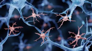
glial cells
Microglia, illustration. Researchers from the New York Genome Center and Columbia University used the atlas’ cross-tissue methylation data to show that a substantial fraction of the brain’s resident immune cells (microglia) are replaced by cells resembling blood monocytes between roughly ages 50 and 75. The finding challenges the long-held view that microglia persist from embryonic development throughout the life span. [Artur Plawgo/Getty Images]

What happens when two epigenetic lenses disagree?

One of the study’s most surprising findings is that DNA methylation and 3D genome structure don’t always tell the same story about a cell. In skeletal muscle, the team found fibers that look like mature, differentiated muscle cells by their 3D genome folding, but still carry the methylation signature of muscle stem cells. The reverse almost never happens. The most plausible explanation, they explained, is that these cells are caught mid-differentiation, with 3D architecture updating first and methylation catching up.

Similar mismatches appeared in Schwann cells of the peripheral nervous system and in placental trophoblasts. The pattern suggests that different epigenetic features update on different time scales during cell state transitions—a finding that could reshape how researchers define “cell type” in adult tissues and how they track cells moving between states in disease.

The atlas also revises a long-standing assumption about “non-CG methylation,” an unusual form of methylation previously thought to be largely confined to brain cells and stem cells. The study shows that it carries cell-identity information across many human tissues, including muscle, pancreas, and immune cell types, at lower but biologically meaningful levels.

“The inconsistency between modalities may be further used to determine what cell populations are switching between each other in adult tissues and diseases, which could, for example, expand our understanding of cancer cell dynamics,” said co-first and co-corresponding author Jingtian Zhou, PhD, a former graduate researcher in Ecker’s lab who now leads his own lab at the Arc Institute.

A public resource for scientists and artificial intelligence

To make the atlas broadly usable, the team built an interactive web browser that lets researchers visualize DNA methylation and 3D chromatin contacts across every tissue, cell type, and subtype in the study. The underlying data, including 195 billion methylation measurements and 18 billion chromatin contacts, are freely available.

The resource arrives as artificial intelligence tools are increasingly used to predict the functional impact of genetic variants. Atlases like this one can provide the labeled, cell-type-resolved training data that models need to make accurate predictions—a bottleneck that has historically limited the field.

For example, in a companion paper in the same issue of Science, a study led by Bing Ren, PhD, from the New York Genome Center and Columbia University used the atlas’ cross-tissue methylation data to show that a substantial fraction of the brain’s resident immune cells, called microglia, are replaced by cells resembling blood monocytes between roughly ages 50 and 75. The finding challenges the long-held view that microglia persist from embryonic development throughout the life span.

“DNA methylation patterns are specific to each cell type and analogous to a cellular barcode,” said Ren, who also co-authored the Salk-led study. “The comprehensive cross-tissue DNA methylation atlases show that the aging microglia in the human hippocampus more closely match the monocytes from peripheral blood than microglia from young adults, providing a crucial clue for the biological identity of these cells.”

The NIH 4D Nucleome consortium, of which this study is a part, aims to extend this kind of mapping into the fourth dimension: time. A 4D understanding of the genome—how its structure and chemistry change as cells develop, age, and respond to disease—remains a major goal, and the cross-tissue atlas provides reference scaffolding that future time-course studies will build on.

Along with scientists from the Salk Institute and Arc Institute,  investigators from the University of California, San Diego, Columbia University, New York Genome Center, University of California, Los Angeles, Harvard, Henan University in China, Vanderbilt University, Stanford University, Broad Institute, University of Sheffield in the U.K., Yale, University of Florida, University of Freiburg in Germany, University of Graz in Austria, and Nanchang University in China; and Chongyuan Luo also contributed to the Science paper.

The post Single-Cell Atlas Simultaneously Maps 3D Genome Architecture and DNA Methylation appeared first on GEN – Genetic Engineering and Biotechnology News.

Midlife Brain Aging Linked to Immune Cell Remodeling, Blood-Brain Barrier Decline

New data from a National Institutes of Health-funded study shows that midlife, the immune cell landscape of the hippocampus, undergoes substantial remodeling. It points to a potential mechanism by which aging may contribute to the chronic neuroinflammation commonly seen in neurodegenerative disease. Details are published in a new Science paper titled “Epigenetic and 3D genome reprogramming during the aging of human hippocampus.”

The work was done by a collaborative team of scientists from the University of California, San Diego, the New York Genome Center, and the University of California, Irvine. According to the paper, the scientists analyzed postmortem hippocampal tissue from 40 neurologically healthy adults aged 20 to 95 years old. 

Digging into the details, the scientists used traditional measures of gene expression alongside more advanced techniques to analyze the genome’s 3D architecture and epigenome. “Gene expression tells us what a cell is doing today, but epigenetic signatures preserve information about where a cell came from,” said Nathan Zemke, PhD, director of single-cell genomics at the UC San Diego Center for Epigenomics and first author on the study. “By combining these approaches, we uncovered a major shift in the identity and lineage of immune cells in the aging human brain’s immune cells that gene expression data alone would not have revealed.”

They found that the brain’s primary immune cells progressively decline from age 50 to 75 years of age, and are replaced by cells with elevated inflammatory signatures and other features that resemble the characteristics of peripheral blood-derived immune cells. It raises questions as to whether microglia, which emerge during embryonic development, may not renew throughout the human lifespan as previously thought. The data also showed that cells that typically maintain the protective blood-brain barrier deteriorated with age. And across many brain cell types, aging accompanied a widespread and coordinated disruption of genome architecture.

“The progressive structural disruptions were closely linked to shifts in gene regulation and cell identity, potentially revealing a fundamental feature of aging in the human brain,” said Bing Ren, PhD, scientific director and CEO of the New York Genome Center, and professor of genetics and development at Columbia University. Ren is also a corresponding author on the study, 

Future studies will investigate the mechanisms driving the loss of resident microglia and determine whether the newly identified immune-cell transition contributes directly to Alzheimer’s disease and other age-related neurological disorders. Insights from the current study as well as others could provide new opportunities to develop therapies that help to preserve brain function and reduce vulnerability to neurodegenerative disease.

The post Midlife Brain Aging Linked to Immune Cell Remodeling, Blood-Brain Barrier Decline appeared first on GEN – Genetic Engineering and Biotechnology News.

Subcortical gray matter atrophy and iron deposition in patients with vascular dementia: a multimodal MRI study

PurposeVascular dementia (VAD) is the second most common type of dementia worldwide. Therefore, early detection and diagnosis, along with a clear understanding of its pathogenesis are critical for mitigating disease progression. In the present study, we aimed to elucidate the associations of brain volume and iron deposition with VAD based on structural brain and iron content analyses.MethodsFifty-three patients with VAD and 43 control participants were recruited for this study. All participants underwent the Mini-Mental State Examination (MMSE) and brain MRI scans. This study primarily focused on the volume of specific brain regions (assessed using FreeSurfer) and iron deposition (evaluated using quantitative susceptibility mapping [QSM]). Linear regression analysis was also performed.ResultsPatients with VAD exhibited significant reductions in brain volume in the left putamen (β = −0.342, 95% CI: −0.581 to −0.104), left pallidum (β = −0.099, 95% CI: −0.187 to −0.001), left hippocampus (β = −0.138, 95% CI: −0.271 to −0.004), and right hippocampus (β = −0.235, 95% CI: −0.420 to −0.051). Additionally, significant increases in iron levels were identified in the left (β = 0.006, 95% CI: 0.002 to 0.010) and right (β = 0.005, 95% CI: 0.001 to 0.009) hippocampus.ConclusionsThese findings indicate that brain volume reduction and increased iron levels in specific regions may be associated with cognitive deficits in patients with VAD.

Memory Shaped by Brain Remodeling During Adolescence in Mice

The human brain continues developing beyond the teenage years, with crucial changes involving decision-making and emotional regulation extending into the mid-to-late 20s. Researchers at Albert Einstein College of Medicine have identified a biological process in mice that offers new insight into how memory circuits mature during this period of brain development. 

The study published in PLOS Biology titled, “Retrosplenial cortical reorganization during late adolescence introduces instability of contextual memory circuits,” found that a key memory region of the mouse brain undergoes a period of remodeling during late adolescence, causing memories formed earlier in life to become temporarily more difficult to retrieve before resurfacing with less precise detail. The findings identify a biological mechanism that may explain how access to memories changes during development. 

The study focused on the retrosplenial cortex (RSP) and discovered that protective mesh-like structures, called perineuronal nets, stabilize memory circuits and unexpectedly diminish during late adolescence before rebuilding in adulthood. The changes were confined to the RSP and were not observed in the nearby hippocampus, another brain region essential for memory. 

“We’ve known for years that the brain continues developing through adolescence and young adulthood,” said senior author Jelena Radulovic, MD, PhD, professor of neuroscience, psychiatry, and behavioral sciences at Einstein. “Our findings begin to explain what that developmental process looks like in one of the brain’s memory circuits and how it can influence the way earlier experiences are recalled. 

Previous studies suggested that the memory circuits reached maturity during early adolescence. Instead, results showed that an important stabilizing system temporarily weakened during late adolescence before recovering in adulthood. 

The timing is notable because it corresponds to a period now recognized as one of continued brain maturation in humans. According to the National Institutes of Health, the brain continues developing and maturing into the mid-to-late 20s. 

“The behavior matched the biology,” said lead author Hui Zhang, PhD, a research fellow at Einstein. “The retrosplenial cortex is responsible for older, more established memories. As its stabilizing structures declined, access to memories formed earlier in life became less reliable.” 

To determine how these brain changes affected behavior, the researchers trained mice to associate a specific environment with a mild foot shock. The mice remembered the experience and froze when returned to the same chamber. Weeks later, many of the mice trained during early adolescence no longer showed that fear response, while mice trained during adulthood retained stable memories over the same period. 

When the adolescent mice later experienced another test in a different environment, they once again responded to the original setting, demonstrating that the memories had become temporarily inaccessible rather than erased. 

The researchers traced these changes to a decline in key structural proteins that help build and maintain perineuronal nets, along with reduced activity of growth factor, TGFβ2. When TGFβ2 activity was restored, the mice regained their ability to retrieve memories formed earlier in life. 

By mid-adulthood, many of those memories resurfaced spontaneously, although they had become less precise. Rather than responding only to the original environment, the mice generalized their fear to unfamiliar settings. The researchers note that this pattern resembles the “reminiscence bump,” a well-known phenomenon in which adults disproportionately recall memories from adolescence and early adulthood while often remembering the emotional significance of an experience more readily than its specific details.  

The findings may also have implications beyond memory. Schizophrenia and major depression often emerge in humans during late adolescence. The authors suggest that changes in this developmental process could contribute to vulnerability to psychiatric disorders in genetically susceptible individuals. Additional research is needed to evaluate whether similar mechanisms occur in humans. 

The post Memory Shaped by Brain Remodeling During Adolescence in Mice appeared first on GEN – Genetic Engineering and Biotechnology News.

Neural mechanisms underlying cognitive inflexibility in obsessive-compulsive disorder: a review

Cognitive inflexibility, a reduced capacity to shift mental set or update behavior when circumstances change, is a significant feature of obsessive-compulsive disorder (OCD). However, while cognitive inflexibility is superficially a coherent entity, it appears to engage a range of distinct cognitive processes. This raises the question of how far deficits in different tasks involve the same core dysfunction and how far the dysfunctions are unique but superficially have similar results. Here we detail the neural basis of OCD deficits across eight tasks that challenge different aspects of cognitive flexibility: set-shifting (Wisconsin Card Sorting Task, Intra/Extra-Dimensional Set Shift), feedback adaptation (Reversal Learning), interference control (Stroop Color and Word Test), response inhibition (Go/No-Go Task and Stop Signal Task), working memory updating (n-back Task), and value-based flexibility (Delay Discounting Task). Cognitive inflexibility in OCD appears linked to functional abnormalities in a largely shared set of core structures (anterior cingulate cortex, caudate nucleus, orbital frontal cortex, and prefrontal cortex) and less shared ‘peripheral’ structures (putamen, thalamus, parietal cortex), based on cross-task convergence of OCD-specific abnormalities. Even the shared core appears to engage multiple interconnected neural networks. The cortico-striato-thalamo-cortical network appears central, and the salience network and the default mode network also contribute with indirect, task-varying effects on peripheral parts of each. Peripheral areas are less consistently involved but appear to contribute to OCD nonetheless. Cognitive inflexibility in OCD appears to involve failure of interaction between multiple networks rather than dysfunction in any single system.

Adolescent exposure to the psychedelic 25C-NBOMe in rats induces lasting competitive avoidance through disrupted hippocampal–prefrontal synchrony

Nature Neuroscience, Published online: 20 July 2026; doi:10.1038/s41593-026-02369-y

Yu, Zhang et al. show that repeated exposure to the psychedelic drug 25C-NBOMe in adolescent rats, but not in adult rats, reduces willingness to engaged in food resource competition in adulthood, due to reduced theta synchrony between ventral hippocampus and orbitofrontal cortex.

Organ Aging Linked to Breakdown in Immune Cell Interaction and Senescent Neutrophil Clearance

We may age at different rates, but none of us escapes aging. A study in mice and in human cells by Stanford Medicine researchers has linked organ aging to the increased inability—with advancing age—of tissue resident macrophage (TRM) immune cells to clear aged neutrophils, another type of immune cell.

The study found that these TRMs appear to be central coordinators of age-related organ decline. Blocking a single receptor, EP2, on these cells preserved the youthfulness of multiple organs in mice, including the brain, heart, skeletal and heart muscle, liver, spleen, bone marrow, kidney, and colon. The receptor binds specifically to a hormone, prostaglandin E2, which is known to cause inflammation and pain in humans as well as in mice.

The researchers found that in mice, selectively disabling this receptor exclusively on tissue-resident macrophages genetically, or using an experimental selective EP2 antagonist drug, prevented chronic-inflammation-driven disorders of age—including frailty, excessive fat accumulation, and heart trouble—and also substantially slowed cognitive decline.

Research lead Katrin Andreasson, MD, the Edward F. and Irene Thiel Pimley Professor in Neurology and Neurological Sciences, said, “We’ve shown that when tissue-resident macrophages don’t have EP2 on their surfaces anymore or when that receptor is plugged up by a drug, this decline doesn’t happen … We’ve been trying to figure out why we age. Now we know at least one big reason for it.”

The discoveries help to clarify systemic inflammation’s significant contribution to aging and the debilities that accompany it. The findings also point to a pharmaceutical approach that could restrain our organs’ unavoidable march toward senescence and so extend overall health span.

Senior author Andreasson, together with first author Jessy Tan, PhD, an instructor in neurology, and colleagues reported on their findings in Science, in a paper titled “Restored clearance of senescent neutrophils by tissue-resident macrophages limits organ aging.” In their research article summary, the team stated, “This work identifies EP2 signaling in TRMs as a central regulator of organ-wide aging through its control of senescent neutrophil clearance, reframing aging as a failure of active cellular clearance rather than passive degeneration.”

Aging is accompanied by parallel functional decline across organs, but the cellular drivers remain unclear, the authors wrote. “Although molecular hallmarks of aging have been identified, the cellular events that initiate and propel tissue decline remain poorly defined.”

The most abundant white blood cells in our immune system are neutrophils, which act as the body’s main first responders. Produced in bone marrow, new neutrophils are transferred to the bloodstream, where they circulate and attack bacterial, viral, or fungal pathogens that they encounter. Neutrophils are also extremely short-lived, surviving just 12–24 hours.

Some 90% of circulating neutrophils end up in the liver, spleen, and bone marrow, awaiting execution clearance by another type of immune cell. “Neutrophils are among the shortest-lived immune cells, aging within hours in the circulation and requiring continuous clearance,” the team noted.

This neutrophil clearance is critical. In aged animals, the vast bulk of neutrophils that never see combat undergo a fast transition to senescence, a zombie-like state in which they may injure, age, and inflame neighboring cells. And as we age, the neutrophil count rises, with senescent neutrophils constituting an ever higher percentage. “Senescent neutrophils are killing our tissues,” Andreasson said. “Clearance of these cells is essential for preventing chronic inflammation.”

That’s a job for macrophages. These cells comb the tissues for pathogens, signal other cells to lend a hand in the fight, and pump out growth factors that help repair damaged tissue. But first and foremost, Andreasson said, “They’re the body’s garbage collection crew. A lot of that garbage is defunct cells.” And a lot of those cells are neutrophils—to the tune of 100 billion a day.

Macrophages come in several subtypes. Tissue-resident macrophages are long-lived and ubiquitous. They take up residence in each of the body’s organs during fetal development and remain for their lifetimes in whatever organ they’ve inhabited, adapting their roles to fit that organ.

One of tissue-resident macrophages’ prime responsibilities is to swallow senescent cells. “A core TRM function is efferocytosis, the clearance of apoptotic, senescent, and damaged cells that is essential for preventing chronic inflammation,” they explained. Especially important targets for this operation, the study showed, are the potentially 100 billion neutrophils produced daily, which start showing signs of senescence within 8 to 12 hours after entering the bloodstream. (Neutrophils that haven’t arrived at senescence yet but have lived long enough and seen enough to put out “kill me now” flags of surrender on their cell surfaces are fair game.)

“Among primary TRM targets are neutrophils, the most abundantly produced immune cell, with more than 10 billion and 100 billion generated daily in mice and humans, respectively,” the investigator noted. “Uncleared aged neutrophils release proteases and extracellular traps that damage tissues, propagate inflammation, and promote aging, and are normally removed efficiently by TRMs in the liver, spleen, and bone marrow.”

But tissue-resident macrophages also grow old. As Andreasson and associates showed in a prior study, over the advancing years these long-lived cells become ever more prone to succumb to aging-associated inflammation and to propagate it. In their newly reported paper, they noted, “TRMs comprise 60–90% of macrophages in the brain, liver, lungs, heart, and kidneys, and their long lifespan makes them particularly vulnerable to aging, as they accumulate metabolic, oxidative, and inflammatory injury over years to decades.”

Immune cells produce hormones called prostaglandins. One of the five varieties of prostaglandin, called PGE2, can exert diverse effects on a cell, depending on which type of surface receptor is expressed on that cell’s surface. Of the various subtypes of receptors for PGE2, the EP2 receptor is highly pro-inflammatory. Tissue-resident macrophages are loaded with EP2.

Infection, injury, and toxic chemicals, including those produced by our aging bodies, increase PGE2 output. As the team’s prior work showed, that output grows substantially as we grow older. So does the concentration of EP2 on tissue-resident macrophages. “TRMs express the prostaglandin E2 (PGE2) receptor EP2, which suppresses macrophage metabolism and phagocytosis in aging,” the investigators noted.

This effectively creates a one-two punch. PGE2’s pro-inflammatory influence increases with age. The resulting unrelenting inflammatory PGE2 stimulation on tissue-resident macrophages, the new study showed, downshifts these cells’ ability to clear neutrophils. Senescent neutrophils then accumulate in tissues and blood.

Andreasson and her colleagues had previously shown that with aging, tissue-resident macrophages undergo a slow decay in their energy metabolism. “Once that starts, there’s a steady decline in a macrophage’s performance,” she said.

For their newly reported study, Andreasson’s lab bioengineered a mouse in which, at a time of the scientists’ choosing, the EP2 gene gets deleted—but only in tissue-resident macrophages. The results of their experiments showed that disappearance of EP2 from these cells reinvigorated the neutrophil-clearance process that PGE2 undermines.

For their experiments, the Stanford Medicine researchers studied younger normal mice, aged 6–8 months, which corresponds to late adolescence or early adulthood in humans, and they also studied older normal mice, at 23 to 25 months of age, whose human counterparts would be in their 60s or 70s. They also looked at older mice whose EP2-encoding gene had been deleted at 4 to 6 months of age (equivalent to their “teenage” years).

The team’s analyses identified 71 proteins, found in blood, whose levels were significantly altered in older normal mice. Of those proteins, 59 stayed at youthful levels in older mice whose tissue-resident macrophages lacked EP2. Many of these proteins originated in the liver. “The liver is one of the body’s most tissue-resident-macrophage-enriched organs and a major contributor to aging-related changes in blood chemistry,” Andreasson said. “It’s the central organ determining the body’s metabolic rate.”

The study showed that in normal old mice, smoldering senescent neutrophils accumulated in the liver, spleen, and bone marrow and, to a lesser extent, in many other organs the researchers looked at.

But the organs of older mice lacking EP2 on their tissue-resident macrophages retained the lower neutrophil numbers of youth. These mice looked younger, leaner, and more physically fit compared with control littermates. They evidenced less visceral fat and greater muscle mass. Their performance on tests of multiple organs’ function equaled that of young mice.

EP2 deletion in addition reduced inflammation in the blood, liver, colon, heart, kidney, and hippocampus (a brain region tightly tied to memory and navigation ability) in the older mice. Their speed, balance, and forelimb grip strength resembled that of young animals.

Reducing EP2 activity in older mice also preserved their memory capabilities. These animals could thread their way through a maze or recall previously encountered objects almost as well as younger mice—and far better than similarly old mice with tissue-resident macrophages expressing functional EP2. “Reducing TRM EP2 signaling in aged mice preserved youthful mitochondrial fitness and prevented cognitive decline, frailty, sarcopenia, adiposity, cardiac impairment, and systemic inflammation,” they wrote in summary.

There are, today, no approved drugs that selectively shut down EP2 activity, although there are several that target PGE2. Non-steroidal anti-inflammatory painkillers work by blocking PGE2 production, Andreasson said. That’s how aspirin and similar drugs reduce pain, fever, swelling, and redness. But to greater or lesser degrees these drugs all block other vital prostaglandins. Even PGE2 has beneficial properties when it binds to receptors other than EP2, rather than the detrimental inflammatory one examined in this study.

As part of their study, the investigators treated otherwise normal 22-month-old mice for two months with an EP2-inhibiting experimental drug. The results showed that the treatment reduced total and senescent neutrophil counts in old mice toward youthful levels. In culture dishes, old age diminished—but the EP2-blocking drug likewise significantly restored—the mice’s tissue-resident macrophages’ ability to engulf and digest burnt-out neutrophils. “Together, these results demonstrate that pharmacologic EP2 inhibition partially reverses age-associated TRM dysfunction and senescent neutrophil accumulation, with strongest rescue in the liver,” they stated.

Finally, the team turned to a large human database characterizing different cell types in young, old, and diseased human livers. This database revealed the same age-related neutrophil buildup, increased neutrophil senescence, tissue-resident-macrophage decline, and heightened EP2 activity in older—and even more so, diseased—livers that the Stanford Medicine researchers had seen in mice. This was a first-time observation in human cells, according to Andreasson. “These human findings, while correlative, position the TRM EP2-efferocytosis axis as a candidate mechanism in human aging that warrants further functional testing,” they noted. “Specifically, future studies should assess whether the impaired clearance of senescent neutrophils also occurs in human TRMs and whether pharmacological EP2 blockade can restore this defect.”

Andreasson suggested that targeting neutrophil clearance may yield big therapeutic benefits. “We need to develop a safe drug that incapacitates EP2 without disrupting upstream events such as PGE2 production.”

The post Organ Aging Linked to Breakdown in Immune Cell Interaction and Senescent Neutrophil Clearance appeared first on GEN – Genetic Engineering and Biotechnology News.

Occupational burnout and risk of suicidality in healthcare professionals: a PRISMA-guided systematic review

BackgroundBurnout, an occupational phenomenon resulting from chronic workplace stress that has not been successfully managed, is increasingly recognized as a critical threat to the mental health of healthcare professionals. Prolonged exposure to work-related stressors may increase the risk of suicidality, including suicidal ideation, suicide attempts, and suicide deaths. This systematic review aimed to synthesize existing evidence on the association between burnout and suicidality in healthcare professionals and to identify vulnerable subgroups and intervention priorities.MethodsWe conducted a systematic review in accordance with Preferred Reporting Items for Systematic Reviews and Meta-Analyses (PRISMA) 2020 guidelines (PROSPERO registration: CRD420251037488). PubMed, Scopus, Web of Science, and PsycINFO were searched for studies published between January 2005 and December 2024. Eligible studies included healthcare professionals and, where methodologically relevant, closely related high-stress occupational populations used as comparator cohorts assessed with validated burnout instruments and reporting suicidality or closely related suicide-proximal psychological outcomes. Data were extracted independently by two reviewers, and risk of bias was evaluated using the Newcastle–Ottawa Scale. Where appropriate, findings were synthesized narratively and through meta-analysis.ResultsA total of 29 studies met the inclusion criteria and 10 studies were included in the meta-analysis. Strong associations were consistently observed between burnout and suicidality, with emotional exhaustion and depersonalization emerging as the most robust predictors. Reduced personal accomplishment demonstrated weaker or inverse associations. Nurses and physicians were identified as particularly vulnerable, with pandemic-era studies reporting higher effect sizes compared to pre-pandemic research. Overall methodological quality was moderate to high, and heterogeneity was partly explained by profession, region, and burnout instrument used.ConclusionsBurnout, particularly emotional exhaustion and depersonalization, is consistently associated with increased suicidality among healthcare professionals, with supporting evidence from related high-stress occupational populations. Vulnerable groups include women clinicians, younger professionals, and those engaged in rotating or night-shift work. These findings highlight the need for systematic burnout surveillance, confidential access to mental health support, and organizational reforms such as safe staffing ratios and workload regulation. Integrating suicide-prevention strategies into occupational health frameworks is urgently required to protect clinician wellbeing and sustain healthcare system resilience.

Dissociation and PTSD in women with a history of childhood sexual abuse: a pilot examination of a specialized inpatient unit

BackgroundWorldwide, there are relatively few specialized inpatient units dedicated to women with histories of childhood sexual abuse (CSA) and comorbid psychiatric disorders. This pilot study examined dissociation and PTSD among women admitted to such a specialized integrative inpatient unit in Israel. We conduct an in-depth analysis of the role of dissociation in these women’s clinical picture, as well as in their treatment response.MethodsThe study included two phases. Phase 1 used a cross-sectional design to assess the complex inter-relationships between PTSD and various facets of dissociation in women with CSA histories admitted to a specialized inpatient unit (N = 108). Phase 2 focused on a sub-sample of participants (N = 28) who completed the inpatient program and completed admission and discharge assessments. Measures included the PTSD Checklist for DSM-5 (PCL-5) and the Dissociative Experiences Scale (DES-II).ResultsIn Phase 1, dissociative symptoms were positively correlated with overall PTSD severity and all PTSD symptom clusters, with the dissociative sub-measure of Absorption showing the strongest associations. In Phase 2, PTSD symptoms significantly decreased following treatment. In line with phase 1, reductions in Absorption were associated with improvements in overall PTSD severity and specifically in Hyperarousal symptoms.ConclusionsFindings from this pilot study indicate the therapeutic potential of a specialized integrated inpatient unit for women with CSA histories. Importantly, our results indicate that dissociation should be regarded as a major therapeutic target, most notably patients’ tendency for maladaptive absorption. These preliminary results should be expanded upon in larger, controlled clinical trials, further elucidating the role of dissociation and other mechanisms of change in similar units.