I spent nearly a decade leading FDA’s Center for Food Safety and Applied Nutrition. That role convinced me that food safety and nutrition are interdependent in so many ways, and if we want to make our population healthier, we need to support both.
Over the past few weeks, record numbers of cases of cyclosporiasis have driven that message home broadly, including to consumers. FDA is currently investigating six cyclospora outbreaks, only one of which is currently linked to shredded iceberg lettuce.
Researchers from University College London in England have created the most comprehensive molecular map to date of severe preeclampsia, uncovering distinct fetal and maternal drivers of the disease that could lead to more targeted therapies and earlier intervention.
The study, published in Science Advances, combined single-cell RNA sequencing with spatial transcriptomics across the entire fetal-maternal interface and found that severe preeclampsia is not solely a placental disorder. Instead, the disease involves coordinated dysfunction across multiple maternal and fetal tissues, with different molecular programs emerging in early- versus late-onset disease.
Preeclampsia affects 2% to 4% of pregnancies worldwide and remains a leading cause of maternal and fetal illness and death. Although it is traditionally viewed as a placental disease, the mechanisms responsible for widespread maternal inflammation and endothelial dysfunction have remained unclear.
To better define those mechanisms, investigators analyzed tissues from 20 pregnancies, including 10 women with severe preeclampsia and 10 gestational age-matched controls between 25 and 37 weeks of gestation. By matching pregnancies by gestational age rather than comparing preterm cases with healthy term pregnancies, the researchers were able to distinguish disease-related molecular changes from normal developmental differences.
The analysis revealed extensive placental abnormalities, including molecular signatures of hypoxia, angiogenic imbalance, fibrosis, and altered metabolism. The team also confirmed impaired invasion of fetal extravillous trophoblasts into the maternal uterus, a hallmark of preeclampsia that contributes to defective remodeling of maternal blood vessels.
Beyond the placenta, however, the study identified previously unrecognized maternal immune abnormalities extending into the myometrium and chorioamniotic membranes. These included widespread mitochondrial dysfunction, activation of type I interferon signaling, and altered macrophage responses, providing a potential explanation for the systemic inflammation and endothelial injury that characterize severe disease.
The findings point to several potential therapeutic targets. Leptin (LEP), which was strongly upregulated in placental cells throughout both early and late disease, may contribute to oxidative stress and vascular dysfunction. The researchers suggest that leptin antagonists, already under investigation for autoimmune diseases, warrant exploration as placental-targeted therapies.
The study also highlights type I interferon signaling as a promising target. Elevated interferon activity, detected in maternal immune cells and peripheral blood, could potentially serve as both a biomarker and a therapeutic target for anti-interferon or antioxidant treatments.
“IFN-I in peripheral blood promises better screening of patients that might benefit from anti-IFN or antioxidant therapeutics,” the authors concluded.
Perhaps most importantly, the molecular abnormalities were substantially more pronounced in early-onset disease, suggesting that intervention before clinical deterioration may offer the greatest benefit.
“Given the severity of molecular dysfunctions in early disease, compared to its late presentation, timely intervention during gestation is likely beneficial and could change the extremely poor prognosis of severe PE,” the authors wrote.
Scientists have identified variants in three genes that influence both the efficacy of CAR T-cell therapy and the likelihood of severe side effects. Published today in Science Immunology, these findings represent a first step toward adopting a precision medicine approach in an increasingly used form of cancer immunotherapy.
CAR T-cell therapies have undeniably transformed the treatment of blood cancers, with a rapidly growing number of approvals over the past decade and progressively expanding toward solid tumors. While genetic variants associated with immune-related side effects have been identified for other forms of cancer immunotherapy, such as checkpoint inhibitors, the role of human genetics in CAR T-cell therapy has remained largely unexplored. However, this is particularly important given that most CAR T cells are manufactured from a patient’s own immune cells, meaning the therapy itself carries the patient’s unique genetic makeup.
“Unlike traditional therapeutics that are essentially identical across all patients, CAR T cells are bespoke and harbor all of the ancestral genetic polymorphisms of their parental T cells,” write the study authors, led by Marcela V. Maus, MD, PhD, professor of medicine at Harvard Medical School and director of the Cellular Immunotherapy Program at Massachusetts General Hospital.
Maus and colleagues analyzed data from 236 patients with aggressive lymphoma enrolled across two clinical trials, who had been treated with axicabtagene ciloleucel, a CAR T-cell therapy commercialized by Gilead under the name Yescarta. The analysis combined whole-genome sequencing with detailed biomarker and functional analysis to identify variants affecting the treatment’s efficacy and toxicity.
Among patients who developed treatment-induced toxicity, many carried variants of the STXBP2 gene. Follow-up experiments in human T cells showed that these variants increased the production of inflammatory cytokines and activated macrophages. Consistent with these findings, mutations in the STXBP2 gene have previously been linked to inflammatory bowel disease and a rare immune disease marked by excessive T cell activation.
In contrast, variants in the ADMTSL3 gene were associated with protection against treatment-related toxicity, while variants in the PTPN22 gene were strongly linked to enhanced CAR T-cell expansion—a critical factor that determines treatment efficacy.
“These findings demonstrate that germline genetics shape the safety and activity of engineered immune cell therapies, affecting future design and patient management,” write the scientists. Going forward, they plan to expand this research into larger patient cohorts and a wider range of CAR T-cell therapies to investigate how different genetic variants influence treatment efficacy and the risk of severe side effects across broader clinical settings.
Beyond opening new avenues for precision medicine in CAR T-cell therapy, the researchers noted that these findings could also inform the development of off-the-shelf CAR T-cell therapies where donor-derived T cells are used instead of a patient’s own cells. This approach could enable a more precise selection of donor T cells optimized to maximize therapeutic benefit while minimizing toxicity risks.
<strong>Background:</strong> Gambling disorder is associated with substantial psychiatric and functional burden, yet few individuals receive treatment. Limited real-world evidence exists evaluating outcomes of virtually delivered behavioral health care for gambling disorder, particularly among clinically complex patients. <strong>Objective:</strong> The purpose of this study was to evaluate gambling symptom severity outcomes among adults with gambling disorder receiving care from Birches Health. We aimed to (1) characterize the clinical profile of adults seeking treatment for gambling disorder; (2) quantify changes in gambling symptom severity over the initial 12 weeks of treatment and examine whether baseline clinical complexity, such as gambling symptom severity, depression severity, and psychiatric comorbidities, was associated with differences in gambling symptom severity improvement over time; and (3) estimate the timing and likelihood of achieving clinically meaningful improvement in gambling symptom severity. <strong>Methods:</strong> This retrospective cohort study included 1305 adults receiving virtual behavioral health treatment for gambling disorder through Birches Health between June 2024 and April 2026. Gambling symptom severity was assessed using the Gambling Symptom Assessment Scale (G-SAS) weekly. Linear mixed-effects models evaluated changes in gambling symptom severity over 12 weeks and associations with baseline clinical characteristics. Clinically meaningful improvement was defined as a reduction of 4 or more points in the G-SAS score. <strong>Results:</strong> Participants had a mean age of 41.5 (SD 13.1) years, 65.2% (851/1305) were male, and baseline gambling symptom severity was moderate (mean G-SAS score 20.5, SD 11.83). Over half (730/1305, 56%) of participants presented with at least one psychiatric comorbidity, most commonly anxiety disorder (351/1305, 26.9%) and depressive disorder (276/1305, 21.1%). Gambling symptom severity declined significantly over the first 12 weeks of treatment, with G-SAS scores decreasing by approximately 0.099 points per day (<i>P</i><.001), corresponding to an estimated 8.3-point reduction over 12 weeks. Higher baseline depressive symptom severity was associated with faster improvement in gambling symptoms (<i>P</i>=.01), whereas depressive disorder (<i>P</i>=.03) and attention-deficit/hyperactivity disorder (<i>P</i>=.008) diagnoses were associated with slower improvement trajectories. Among patients with routine follow-up assessments recorded during the initial 12 weeks of treatment (1071/1305, 82.1%), 71.7% (935/1305) achieved clinically meaningful improvement in gambling symptom severity, with a median time to improvement of 14 days. <strong>Conclusions:</strong> A clinically complex population of adults receiving care through a national virtual behavioral health care provider demonstrated rapid and clinically meaningful reductions in gambling symptom severity. These findings highlight the potential of specialized virtual care models to expand access to gambling treatment and support symptom improvement in routine care settings. Future research should evaluate longer-term recovery trajectories and identify factors associated with sustained improvement and ongoing engagement in care.
WASHINGTON — A Food and Drug Administration advisory panel recommended on Friday that compounding pharmacies be allowed to manufacture the peptides epitalon and semax, but narrowly voted to recommend against manufacturing emideltide.
The votes, which followed the panel’s decision on Thursday to recommend allowing pharmacies to make four other peptides, bring health secretary Robert F. Kennedy Jr. one step closer to his mission of making these unapproved compounds more available for Americans. Peptides, which are short-chain amino acids, have become increasingly popular in the U.S., driven by endorsements from social media influencers.
Researchers at Kobe University have generated off-the-shelf, mass-producible induced pluripotent stem cell (iPSC)-derived gamma delta T cells (γδT cells) that in a small preclinical study suppressed tumor growth in mouse colorectal cancer (CRC) xenograft models. The team says their development could point to the potential for developing faster, cheaper cancer immunotherapy.
“Various immunotherapies have been developed to treat malignant tumors, and autologous CAR T-cell therapy is clinically used for certain malignancies,” the authors wrote. However, CAR T-cell therapies demonstrate limited efficacy against solid tumors, and current techniques for modifying T cells extracted from the patient are expensive and time consuming. “… obstacles such as the time and cost required to initiate autologous treatment impede their widespread adoption.”
In the journal Stem Cell Reports, Kobe University stem cell researcher Aoi Takashi and his team report that they created iPS cells from a subclass of T cells that can be used across patients and could reproducibly turn them back into T cells with an overall 80,000-fold multiplication and without using animal cells or extracts, and that the resulting T cells attack and shrink human patient-derived colorectal cancer tumors that were implanted into mice. [Aoi Takashi]
Consequently, the authors noted, there is growing interest in allogeneic, or off-the-shelf, cell therapy. Researchers have considered turning to a subclass of T cells called gamma-delta (γδ) T cells that don’t need to be tailored toward each individual patient but can be harvested from a donor and used in other people. “… the development of novel therapies for CRC, a highly heterogeneous cancer, remains a paramount challenge in global healthcare, and γδT cells are considered a promising candidate modality,” the authors stated. “γδT cells represent approximately 3–5% of peripheral blood lymphocytes and are capable of targeting various types of tumors in an MHC-unrestricted manner with a single type of γδT cell receptor.”
However, these cells are much fewer, making the harvesting approach infeasible, and they also cannot be directly multiplied well in the lab. Aoi stated, “Based on our experience with induced pluripotent stem cells, also called iPS cells, we thought that we could approach this issue by creating such easily storable and growable cells from these specific T cells, and then only turning them back into T cells when actually needed.” In their paper the authors added “We focused on γδT cells as a potential therapeutic modality for colorectal cancer (CRC).”
Through their reported study the investigators showed that they could create iPS cells from the subclass of T cells that can be used across patients and reproducibly turn them back into those T cells with an overall 80,000-fold multiplication. Importantly, they achieved this without relying on animal cells or extracts, which is a requirement for clinical applications. “To the best of our knowledge, this is the first study to report the successful induction of differentiation of γδT cells from iPS cells under feeder-free, serum-free conditions.”
Their study was also the first to show, on a small preclinical scale, that the resulting iPSC-derived γδT cells (iγδT cells) attack and shrink human patient-derived colorectal cancer tumors that were implanted into mice, with tumor weights in treated animals reduced by up to 88%, when compared with control mice.
“We demonstrated that these iγδTs exhibit cytotoxic activity against CRC and leukemia cell lines, as well as against patient-derived CRC organoids in vitro, while also exerting antitumor effects in vivo in xenograft models,” they noted. “Cancers from cell culture lines don’t have the same drug insensitivities as actual cancers and also don’t emulate the physical barriers that actual tumors have,” explained first author Ryoko Futai, PhD. “That’s why patient-derived organoids are highly significant for evaluating new cancer treatment approaches,” explained first author.
When they designed the study, the Kobe University team imagined that their approach would be used fighting metastasizing cancers. They also checked whether their T cells would find their targets not only when administered close to the tumor but when administered intravenously a week after the tumor was implanted. And indeed, even in this setting tumor weights decreased 43%, 82% and 92% in the three treated mice. Futai noted, “This suggests potential for future systemic therapy. We believe this achievement represents an important step toward the development of a new immunotherapy for solid tumors.”
The study was conducted at a small scale, with only three or four mice in each experiment and tumor models derived from only two different patients. This is especially important because colorectal cancer tumors are known for their high variability. “This study is a preclinical investigation demonstrating the potential using iPS cell-derived T cells and is not yet at a stage where it can be used on patients,” cautions Futai.
But by conducting further studies using these easily multipliable and very standardized cells, the Kobe University development may also be used to elucidate where the variability comes from and what steps to take to counter it. Aoi commented, “Furthermore, by combining this approach with cell modification techniques such as CAR therapy, we hope that this research will eventually lead to the development of new therapeutic possibilities for patients with solid tumors.” And in their paper the authors concluded, “Our findings will pave the way for the realization of off-the-shelf allogeneic γδT cell therapy.”
This week, I covered a fascinating effort to preserve organs outside the body. There’s a huge shortage of donor organs, and one of the main reasons is time—they survive only a matter of hours outside the body, even when they’re kept on ice.
Doctors dream of organ banks—stores of human organs that can be preserved for days, weeks, months, or even longer. That would allow them to run tests on organs, find the best matches for them, and transport the organs to those recipients.
In new research, one team has been able to supercool the kidneys of pigs—animals whose organs are of a similar size to human ones—and preserve them for days. The kidneys survived being stored at −4 °C (25 °F) and eventually reimplanted back into pigs. And that’s just the latest development in a field that is positively buzzing.
It has proved super difficult to freeze organs. Once ice forms in them, they’re done. The ice crystals create all kinds of damage and render the organs unusable. That hasn’t stopped many researchers from trying.
Some have focused on cryopreservation—rapid extreme cooling that essentially leaves cells in a glasslike state. This process is now routine for eggs, sperm, and embryos, which are cooled to −196 °C in less than two seconds and can be used even after decades in storage.
No one has managed to cryopreserve and thaw human organs for transplantation. But plenty of human bodies and brains have been stored at ultra-low temperatures in the hope that they might one day be rewarmed and brought back to life. (You can read more about why some people opt for cryonics here.)
In March, I wrote about Stephen L. Coles, a gerontologist who had opted to cryopreserve his own brain. After the scientist died in 2014, his body was taken to Alcor, a cryonics facility in Arizona. A team at the facility removed Coles’s head, perfused his brain with cryoprotective chemicals (which work like antifreeze), removed the brain from the skull, and cooled it to −146 °C.
When Coles’s friend Greg Fahy, a cryobiologist, studied pieces of his brain years later, he found that the brain cells, which had shrunk, “bounced back” once they were rewarmed. But that doesn’t mean the cells are alive, or that it might one day be possible to reanimate the brain. As Matthew Powell Palm of Texas A&M told me at the time: “There are so many ways those neurons could be toast.”
Powell Palm is working on other ways to preserve organs. It was he, along with his colleagues, who managed to store supercooled pig kidneys and successfully transplant them, in a study described as “a landmark achievement.” Those organs did better than kidneys stored on ice, he says.
His approach didn’t require cryoprotectants. But other teams are exploring potential chemical cocktails that might allow them to store organs at lower temperatures, potentially for longer periods of time. (More on this in The Checkup soon!)
Another way to prolong the lifespan of an organ is to use a machine that perfuses it with nutrients, mimicking what happens inside the body. Machine perfusion devices have become more commonly used over the last decade or so and are typically used to maintain livers and kidneys for up to about 24 hours.
Researchers are now adapting this protocol for a growing list of organs, even eyeballs—a recent feat that might enable whole-eye transplants. In March, I went to visit scientists in Valencia who had developed a perfusion system for uteruses. They had used their device—which they nicknamed “Mother”—to keep a human uterus alive for a day.
It’s an exciting time for organ preservation. Keep an eye out for more coverage from MIT Technology Review in the coming weeks.
This article first appeared in The Checkup, MIT Technology Review’s weekly biotech newsletter. To receive it in your inbox every Thursday, and read articles like this first, sign up here.
Posted on
<![CDATA[New BEST teletraining helps people after TBI name emotions, reduce anxiety and anger, and boost resilience—early trials show promise.]]>
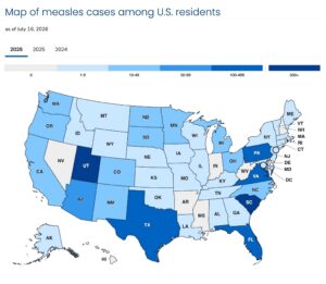
This year, the United States has recorded the highest number of measles cases since 2000, the year that the disease was declared eliminated from the country. As of mid-July, 2,260 measles cases were reported—just 29 cases less than the entire year of 2025. And outbreaks are widespread, with 34 new outbreaks reported in 2026.
The reason for this surge in cases is waning vaccination rates across the country. Given that, the production of new antivirals is an urgent matter.
Map of measles cases in the U.S. [CDC]
Now, the new oral antiviral drug candidate GHP-88310 has shown promising results in a ferret model of infection. When administered before or after direct contact or airborne exposure to canine distemper virus (which causes measles-like disease in ferrets) GHP-88310 blocked transmission of the virus and reduced clinical symptoms in ferrets.
“We were very excited to see that GHP-88310 given by mouth completely prevented airborne transmission in our ferret model of measles,” said Carolin Lieber, PhD, a postdoctoral fellow in the Plemper lab at Georgia State University. “This finding is unprecedented for a viral polymerase inhibitor and demonstrates the extraordinary antiviral potency of this drug.”
“Silencing measles outbreaks quickly is essential to reestablish control over the virus,” said Richard Plemper, PhD, professor and director of the Center for Translational Antiviral Research (CTAR) at Georgia State University. “This study follows our recent development of the drug candidate GHP-88310. It demonstrates that the drug is suitable to augment traditional ring vaccination against measles.”
GHP-88310 (described earlier this year in Science Advances) is known to be an orally efficacious broad-spectrum orthoparamyxovirus polymerase inhibitor. But its effect on viral transmission has remained unclear. This study explored whether prophylactic administration of GHP-88310 prevents virus transmission through close contact or through the air. The results demonstrate that GHP-88310 efficiently blocks both forms of viral spread. In addition, the study showed that treatment of infected animals shortened the time period in which infected animals could transmit the virus.
To explore relevant conditions of viral transmission, the researchers established both direct-contact and airborne canine distemper virus transmission models to examine pharmacological suppression of virus spread. The transmission systems allowed them to pair infected and uninfected animals in direct physical contact or shared airspace, each under controlled environmental parameters.
“We designed the study to recapitulate viral spread between people with direct contact, for instance in a household, and between more distant social contacts, for example in classrooms or other indoor settings that bring people into proximity without direct interaction,” said Plemper.
The results showed that pre- and post-exposure prophylactic GHP-88310, given twice daily to air contacts, prevented transmission. The authors note that once-daily prophylactic administration mediated complete survival with all air contacts undergoing seroconversion. In addition, they note that therapeutic treatment of air contacts mitigated clinical signs, and animals survived, whereas all vehicle-treated air contacts succumbed. In addition, therapeutic treatment of infected source animals shortened the contagious phase by five days.
“In addition to this prophylactic benefit, GHP-88310 used therapeutically shortened the duration of disease in our model,” noted Plemper. “If equally applicable to human hosts, it may shorten the severe social and economic burden of prolonged quarantine of patients and further aid outbreak management.” The investigators are now readying GHP-88310 for formal clinical testing.
A chemical called trimethylamine N-oxide (TMAO), which originates from gut bacteria digesting animal products such as red meat, is present at higher levels in the blood of people with an irregular heart beat than the rest of the population.
The researchers found that after they had controlled for various factors like age and comorbidities, people in the top third of TMAO levels had a 70% increased risk of having atrial fibrillation compared with those in the bottom third.
Atrial fibrillation is the most common sustained heart rhythm disorder in the U.S. impacting more than 10 million adults. It occurs because the upper chambers of the heart beat rapidly and irregularly, which can lead to blood clots, stroke and heart failure if left unchecked.
TMAO is formed when trimethylamine produced by gut bacteria is carried to the liver and oxidized. Animal-derived foods such as red and processed meat, eggs, high fat dairy products, and some supplements, are the main dietary precursors for production of TMAO. It has previously been linked to increased risk of cardiovascular disease more generally, but whether high levels of this chemical increased risk of atrial fibrillation was less clear.
In this study, published in the journal of Journal of Clinical Investigation, lead investigator Robert Koeth, MD, PhD, a clinician scientist based at the Cleveland Clinic, and colleagues assessed blood TMAO levels in 5,000 adults who underwent elective heart catheterization for known or suspected cardiovascular disease.
They also studied the effects of TMAO on heart beat regularity in model mice susceptible to atrial fibrillation and tested a potential candidate drug to see if it could reduce levels of TMAO in these animals.
After controlling for age, sex, diabetes, cardiovascular disease, blood pressure, smoking, body mass index, inflammation and kidney function, people with the highest TMAO levels had about 1.7‑times the odds of having atrial fibrillation compared with people with the lowest levels.
In the mouse studies, when TMAO levels in the blood were higher, the animals developed atrial fibrillation sooner and it became a persistent problem more quickly. The researchers also saw changes in how electrical signals moved through parts of the heart that made it easier for abnormal rhythms to start and continue.
The researchers tested iodomethylcholine in the mice, an experimental small molecule drug that blocks the gut microbial enzyme responsible for converting choline into trimethylamine. In response to the drug, TMAO levels fell, the mix of gut bacteria changed, and both the first episodes of atrial fibrillation and its progression were delayed without notable negative effects on the liver.
“A possible pathogenic culprit for the development of atrial myopathy and electromechanical dysfunction is chronic autonomic dysfunction from TMAO inhibition of the M2 muscarinic acetylcholine receptor and increasing sympathetic tone,” write Koeth and team.
The results suggest “TMAO promotes atrial fibrillation in a gut microbiota dependent manner by causing autonomic dysfunction, atrial myopathy, and electromechanical dysfunction,” they conclude.



![In the journal Stem Cell Reports, Kobe University stem cell researcher AOI Takashi and his team report that they created iPS cells from a subclass of T cells that can be used across patients and could reproducibly turn them back into T cells with an overall 80,000-fold multiplication and without using animal cells or extracts, and that the resulting T cells attack and shrink human patient-derived colorectal cancer tumors that were implanted into mice. [AOI Takashi]](https://www.genengnews.com/wp-content/uploads/2026/07/Low-Res_260723-Aoi-CRC-Incubator-300x233.jpg)