

Transmissible Cancer Discovered in Freshwater Fish

Researchers headed by a team at the University of Vermont have discovered that mysterious black skin lesions afflicting catfish in Lake Memphremagog and other New England and Canadian lakes are caused by a transmissible form of cancer, the first ever identified in a freshwater fish species. The scientists’ studies showed that the cancer cells behave more like parasites than conventional tumors, moving from fish to fish.

The discovery marks only the fourth type of transmissible cancer identified in the animal kingdom, and the first in any fish or freshwater species. Previous ones include the Tasmanian devil’s facial tumor disease, a transmissible venereal tumor in dogs, and transmissible leukemia-like diseases in clams, mussels, and other bivalve mollusks. The researchers do emphasize that the disease poses no known risk to humans. The cancer cells cannot infect or survive in another species, and the fish are safe to handle and study.

The new discovery sheds light on how cancer can spread in the wild, and raises important questions about where this cancer originated, how it will affect fish health and populations, and also how cancer works in all animals including humans.

Julie Dragon, PhD, a researcher at the UVM Cancer Center in the Larner College of Medicine, suggests that understanding this newly discovered transmissible cancer could offer new insights into cancer biology more broadly. “By studying how cancers survive and spread outside their original host, we can learn a great deal about what keeps cancers contained—and what happens when those boundaries break down,” she said. “These fish provide an opportunity to look at evolution of cancer.”

Dragon is co-lead author of the researchers’ published report in Nature, titled “Brown bullhead catfish melanoma represents a novel transmissible cancer,” in which they stated, “We found extensive genomic evidence to suggest these melanistic lesions represent a transmissible cancer, to our knowledge, the first known occurrence in a fish species and the first found in a freshwater aquatic setting.”

Since 2012, anglers and biologists have reported a striking increase in brown bullhead catfish (Ameiurus nebulosus) with raised black skin patches in the cross-border lake shared by Vermont and Quebec. By 2014, nearly one in three fish showed the dark lesions. Because brown bullhead are considered indicators of environmental quality, the researchers suspected a contaminant or pathogen linked to pollution. “At first, we thought this disease might be a virus, but that wasn’t panning out,” said Dragon.

Instead, scientists found that these lesions turned out to be melanoma, a skin cancer. “This was surprising,” Dragon said, “and we wanted to know how a bottom-dwelling fish was getting a cancer we associate with exposure to too much sunlight.” For their reported study Dragon and colleagues took what she describes as a “deep dive,” into the genetics of the cancer cells.
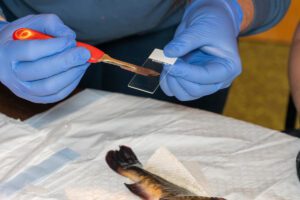
UVM professor and cancer researcher Julie Dragon co-leads dissection and tissue collection of catfish with melanoma skin cancer at the Gateway Center on the shore of Lake Memphremagog, Newport, Vermont. Additional participants: Elizabeth Murchison, professor at the University of Cambridge, UK; her post-doctoral associate Zoe Clarke; USGS post-doctoral research fish health biologist Cheyenne Smith; her undergraduate student intern Sam Williams (not shown in these photos); Peter Emerson, fish biologist with the Vermont Department of Fish & Wildlife. The team sampled tissue from 17 fish.
UVM professor and cancer researcher Julie Dragon co-leads dissection and tissue collection of catfish with melanoma skin cancer at the Gateway Center on the shore of Lake Memphremagog, Newport, Vermont. Additional participants: Elizabeth Murchison, professor at the University of Cambridge, U.K.; her post-doctoral associate Zoe Clarke; USGS post-doctoral research fish health biologist Cheyenne Smith; her undergraduate student intern Sam Williams (not shown in these photos); Peter Emerson, fish biologist with the Vermont Department of Fish & Wildlife. The team sampled tissue from 17 fish. [University of Vermont]

Using whole-genome sequencing, the team compared DNA from tumors and healthy tissues in affected fish. They expected to find a genetic mutation that makes the fish susceptible to having their own cells become cancerous. Instead, they found that the cancer cells were much more closely related to each other than to their host fish—the signature of a clonally transmissible cancer, in which tumor cells themselves act as infectious agents.

“Hundreds of thousands of genetic variants are shared among tumor samples but absent from host fish, vastly exceeding levels seen in conventional cancers,” the authors reported in their paper.

The research team is now working to understand how the cancer cells spread between animals. “It seems to only happen in larger fish that are of spawning age,” said study co-lead Mark Henderson, PhD, fish biologist at UVM’s Rubenstein School of Environment and Natural Resources. “Maybe some part of spawning behavior leads to the spreading of the cancer between animals.”

The study notes that pollutants or hormonal changes could weaken immune systems, making the lake’s brown bullhead fish more vulnerable to infection. Naturally occurring arsenic may also play a role. The team did find that the fish cancer cells have elevated levels of arsenic and the geographic distribution of the cancer appears to have correlation with levels of arsenic in the lakes’ surrounding soil. “Arsenic is found naturally throughout New England, and there’s really high concentrations of it in the Vermont’s Northeast Kingdom as well as up through Maine, which is where we’ve also seen evidence of this cancer,” said Henderson.

The cancer’s ecological impact remains uncertain. Some transmissible cancers are devastating—the Tasmanian devil tumor has wiped out 90% of some populations. In contrast, the dog tumor has coexisted with its hosts and has done for thousands of years. In Lake Memphremagog, heavily diseased bullhead can survive for years, but long-term population effects are still unknown.

The researchers are beginning to explore how long this cancer may have been around, and how common transmissible cancers may be. They are exploring ponds and rivers in Massachusetts to learn more about the distribution of the cancer and to see if it may have originated there. “Our contention is that maybe it’s not as rare as everyone thinks. We’re just not seeing it,” said Dragon. “In the case of transmissible cancers, it may be that we don’t see many simply because we aren’t looking for them.”

In their paper the team concluded, “Although we cannot rule out a role for conventional pathogens in the original emergence or ecological transmission of this disease, the genomic evidence presented here overwhelmingly supports a transmissible cancer in which the tumor cells themselves act as the infectious entity.”

The post Transmissible Cancer Discovered in Freshwater Fish appeared first on GEN – Genetic Engineering and Biotechnology News.

Breath Sensor Monitors Fat Metabolism at Home

Scientists in Switzerland have developed a portable breath detector that can accurately measure acetone released into the breath when the body burns fat. The smartphone-assisted device allows patients to monitor their metabolism at home and could help doctors personalize treatment for metabolic diseases such as obesity and diabetes.

Acetone levels in the breath have long been recognized as an indicator of metabolic activity, since its concentration rises when the body shifts from using carbohydrates to fats as the primary energy source. However, accurate measurements have traditionally required bulky and expensive laboratory equipment, while consumer devices have lacked the sensitivity needed to reliably measure acetone, especially at lower concentrations. 

“If we want to make that information available to patients, we need to shrink those technologies into compact, user-friendly devices,” said Andreas Güntner, PhD, assistant professor at ETH Zurich and senior author of the study published today in the Device journal.

To make a compact breath analyzer, Güntner’s team used a chemoresistive sensor that changes its electrical properties in the presence of acetone. These types of sensors are known for their high sensitivity, rapid response, low power consumption, and small size. During each measurement, the accompanying smartphone app coaches users to exhale with the right force and duration, while quality controls can reject improper breaths or contaminated air. This design makes the sensor easy to use while ensuring accurate measurements. 

The breath detector was used to analyze 312 breath samples from 12 healthy adults, with measurements closely matching those obtained using gold-standard mass spectrometry. “These findings show that we have the high performance needed for applications such as clinical studies, where you really want to distinguish these slight differences in fat metabolism,” said Simone Hersberger, graduate student at ETH Zurich and first author of the study. 

The researchers then used the sensor to monitor breath acetone under four metabolic scenarios: light exercise followed by a high-carbohydrate meal, intense exercise followed by a high-carbohydrate meal, a high-fat ketogenic meal, and fasting. Breath acetone levels remained low during light exercise but increased with intense exercise, dropping after a high-carbohydrate meal. Acetone levels rose after a ketogenic meal and increased further during fasting. 

Through Alivion, a spin-off from ETH Zurich, the technology is already available to individuals interested in tracking breath acetone to monitor weight loss and athletic performance. The device is currently being used to monitor individual progress in clinical studies of epilepsy, where a ketogenic diet is a standard medical treatment. 

“Now it’s really time to spread it out into clinical trials and answer questions such as the effectiveness of different fasting therapies by providing personalized guidance,” said Güntner. “We’re really moving toward healthcare solutions that, in the future, you won’t need to go to the hospital for anymore. You’ll be able to do them at home.” 

The post Breath Sensor Monitors Fat Metabolism at Home appeared first on Inside Precision Medicine.

Therapy‑Driven DNA Changes in Pediatric Tumors Can Spur Resistance

Research led by the Hospital for Sick Children in Toronto shows chemotherapy and radiotherapy are key sources of mutations in relapsed childhood tumors and that different treatments leave distinct mutational signatures.

As described in Nature, across all mutations in these pediatric tumors, about 15% can be clearly traced to four chemotherapies, and most of that therapy‑linked damage comes from platinum drugs like cisplatin, carboplatin, and oxaliplatin.

After platinum chemotherapy, temozolomide, 5‑FU, and thiopurines were the next‑most clearly mutation‑linked drugs in this study, but each added only a small fraction of the total mutational burden compared with platinum drugs.

“Many of the drugs used to treat children with cancer cause unfortunate long term side effects, including heart issues and secondary cancers,” lead author Adam Shlien, PhD, told Inside Precision Medicine.

“These drugs can also lead to somatic mutations, although the total genomic burden of this wasn’t known. Whether these mutations are associated with drug resistance in childhood cancer was also mostly unknown.”

In this study, the researchers assembled a multi‑national precision‑oncology cohort of 611 tumors from 544 children and young adults enrolled in three whole genome sequencing‑based programs, focusing on aggressive, relapsed or metastatic cancer.

The team then looked at exposure to 86 types of therapy in 13 drug classes, as well as radiotherapy, and created a detailed record of cycle dates, doses, and routes of administration for each child. They also recorded the number of drugs or other treatments the cancer patients were exposed to, looked for mutation patterns in tumor DNA linked to specific chemotherapy agents and tracked when these first appeared after treatment.

“It was striking how many mutations are associated with therapy—when the tumor cells survive, they frequently acquire thousands of mutations and many of these are tightly linked to the type of therapy that was used,” explains Shlien.

Platinum drugs were the biggest contributors to tumor mutation signatures. They caused a large fraction of all therapy‑related mutations and left clear, characteristic mutation patterns that appeared in a short amount of time after starting treatment, sometimes in as little as three months.

Although the presence of mutations did not necessarily lead to drug resistance or relapse, tumors with strong platinum‑linked patterns often showed activation of genes known to help cancer cells resist treatment with platinum drugs.

This finding was confirmed in the pediatric study cohort and in adults with cancer treated with platinum chemotherapy. Patients whose tumors carried these patterns had worse outcomes when treated with platinum drugs.

This study opens the door to using these mutation patterns to guide care. This could include deciding when to avoid re‑using a drug, or when to consider lowering doses of mutagenic treatments like platinum chemotherapies in settings where cure rates are already high. It also shows that these treatment‑induced mutation patterns are not just signs of past therapy but can be an early warning of the emergence of drug‑resistant cancer cells.

“Now that we have comprehensively defined which therapy-associated mutation patterns are acquired in childhood cancer, and when they emerge, we can start to think about screening patients for these signatures for the early detection of drug-resistant clones,” says Shlien.

The post Therapy‑Driven DNA Changes in Pediatric Tumors Can Spur Resistance appeared first on Inside Precision Medicine.

Malaria Drug Reveals Genetic Vulnerability Across Cancers

Scientists have found that the antimalarial drug quinacrine can exploit a vulnerability in cancers that become reliant on the NDRG1 protein to sustain their DNA damage response. Published in Science Signaling, their study not only identifies NDRG1 as a promising therapeutic target, but also demonstrates a new strategy for discovering similar vulnerabilities across multiple types of cancer.

“Our findings suggest that NDRG1 expression could serve as a biomarker to help identify patients most likely to benefit from therapies targeting this pathway,” said Garik V. Mkrtchyan, PhD, assistant professor at the University of Copenhagen and lead author of the study. “We further showed that high NDRG1 expression predicts poor survival across multiple cancers, and that inhibiting NDRG1 creates vulnerabilities, highlighting new opportunities for precision oncology.”

Mkrtchyan and colleagues set out to identify new targets for cancer therapeutics by leveraging the concept of synthetic lethality—a phenomenon where cancer cells can survive the loss of one of two genes but die when both are inhibited. This approach has already proven successful in ovarian, breast, and prostate cancers with BRCA mutations, which are particularly vulnerable to PARP inhibitor drugs. 

“In oncology, this concept is particularly promising because cancer cells often harbor mutations in specific DNA damage response pathways, making them highly dependent on the remaining repair mechanisms for survival,” said Mkrtchyan. “Targeting these dependencies enables selective elimination of cancer cells while sparing healthy tissue.”

Using transcriptomics data, the researchers identified quinacrine as a promising candidate for disrupting the DNA damage response by targeting the stress-response protein NDRG1. Quinacrine has been used as an antimalarial drug for nearly a century, later gaining approval as a treatment for lupus. In recent years, the compound has attracted growing interest as a potential cancer treatment. 

Screening through hundreds of cancer cell lines revealed that blood cancers, which generally showed high NDRG1 expression, were the most sensitive to the drug. Colorectal cancer cells were also sensitive to quinacrine, especially those with mutations in the MLH1 and PARP3 genes. The team later confirmed these findings in patient datasets, where high NDRG1 expression together with loss of either of these genes correlated with improved survival rates. 

“While quinacrine has previously been reported to possess anticancer activity, our study uncovers upstream mechanisms of its action on DNA damage response,” said Mkrtchyan. “By applying an automated robotics screen across more than 130 cancer cell lines, we identified novel synthetic lethal interactions involving NDRG1, providing a framework for discovering new therapeutic vulnerabilities across multiple cancer types.”

Despite its potential as a cancer therapy, quinacrine can potentially cause unwanted side effects. The researchers therefore plan to explore alternative drug candidates that can inhibit NDRG1 with more potency while reducing toxicity.

“The next steps will be to develop small molecules that inhibit NDRG1 with greater potency and specificity than quinacrine, thereby minimizing potential off-target effects,” said Mkrtchyan. “From a translational perspective, we aim to validate the identified synthetic lethal interactions in preclinical tumor models and investigate whether targeting the NDRG1 axis can overcome treatment resistance across a broader range of cancers.”

The post Malaria Drug Reveals Genetic Vulnerability Across Cancers appeared first on Inside Precision Medicine.

ctDNA Blood Test Better Targets Radiopharmaceutical Prostate Cancer Therapy

A new ctDNA blood test from a Japanese team identifies those metastatic, castration-resistant prostate cancer (mCRPC) patients most likely to benefit from 223Ra radiopharmaceutical therapy. The test also monitors their progress throughout treatment.

The study appeared in the July issue of The Journal of Nuclear Medicine. The lead author is Masaki Shiota, MD, PhD, associate professor in the Department of Urology in the Graduate School of Medical Sciences at Kyushu University in Fukuoka, Japan. 

Prostate cancer is very biologically heterogeneous. Patients who appear clinically similar may have different responses. Better tests are needed to match patients to therapies and determine the optimal treatment sequence or combination of therapies.

223Ra dichloride targets bone metastases and can improve overall survival and quality of life in CRPC patients. It is an alpha-particle–emitting radionuclide that behaves chemically like calcium. It is therefore preferentially incorporated into areas of increased bone turnover, including the osteoblastic lesions commonly produced by prostate cancer bone metastases.

But the treatment is expensive, clinical outcomes vary among patients, and there is not yet a reliable biomarker to predict or monitor treatment response.

This study used a research-based 88-gene panel with deep sequencing of plasma circulating tumor DNA (ctDNA) and matched leukocyte DNA. “The leukocyte analysis was important for distinguishing true tumor-derived alterations from germline variants and age-related clonal hematopoiesis,” Shiota told Inside Precision Medicine.  

Prostate cancer is one of the most commonly diagnosed malignancies in men. Metastatic castration-sensitive prostate cancer arises either de novo as a cancer, or as a recurrence after radical local therapy for localized disease. It accounts for approximately 10% of newly diagnosed prostate cancer cases in Japan.

This team tracked associations between ctDNA profiles and clinical outcomes, including biomarker response, radiographic progression-free survival, and overall survival. 

Patients with a higher amount of tumor DNA in the blood or certain gene changes, such as TP53, PTEN, and cell cycle pathway alterations, detected through ctDNA testing before treatment, had worse outcomes. Further, changes in tumor DNA reflected treatment response and disease trajectory.

“While 223Ra is an important treatment for prostate cancer that has spread to the bones, not all patients benefit equally,” said Shiota. “Our findings suggest that a blood-based genomic test may help identify patients who are more likely or less likely to benefit from the therapy. This could help doctors choose treatment more carefully and monitor patients more closely, with the goal of providing more personalized care.”

One of the biggest hurdles to the test’s advancement is reimbursement.

This depends on the country, the clinical indication, and the specific assay used. In Japan, Shiota explained, certain approved comprehensive genomic profiling tests using blood, such as FoundationOne Liquid CDx, can be covered by the national health insurance system under defined conditions. These tests are primarily used to identify potentially actionable alterations in patients with advanced solid tumors, particularly when obtaining adequate tumor tissue is difficult.

“The research assay used in our study is not currently reimbursed for selecting patients for radium-223 or for monitoring their response during treatment,” said Shiota. “Before insurance coverage could be considered for this particular use, our findings would need to be externally validated, and prospective studies would need to demonstrate that ctDNA-guided treatment decisions improve patient outcomes.”

ctDNA has several important applications in precision oncology. For example, it can: identify actionable genomic alterations and thereby help select targeted therapies, be used to monitor treatment response and detect emerging resistance mechanisms, capture tumor heterogeneity more comprehensively than a biopsy from a single metastatic site, detect minimal residual disease after treatment, and identify molecular relapse before it becomes apparent on conventional imaging.

The post ctDNA Blood Test Better Targets Radiopharmaceutical Prostate Cancer Therapy appeared first on Inside Precision Medicine.

Transcripta Bio Raises $24M for AI-Driven Neurological Disease Therapies

Chris Moxham, PhD, has been a “drug hunter” for three decades, building data-driven platforms as vice president of quantitative biology at Eli Lilly, and CSO of Folcrum Therapeutics, in recent years. 

“Technology is now advancing to allow us to interrogate the transcriptome, which is a phenomenal blueprint for cell state and fate,” he told GEN Edge.  

Moxham currently leads Transcripta Bio as founder and CEO. The AI-driven drug discovery start-up is developing small molecule therapeutics to modulate gene expression disease signatures. 

Transcripta has now announced a $24 million funding raise to advance IND-enabling studies and clinical preparation of its neurological disease portfolio, including autism spectrum disorder (ASD) and facioscapulohumeral muscular dystrophy (FSHD). Mayo Clinic and Omnimed will join JAZZ Venture Partners, BlueYard Capital, and a group of life sciences family offices, as investors. 

Phase II clinical trials are “where the rubber meets the road,” says Moxham. He emphasizes that de-risking therapies early is key to improving drug discovery success rates, which often fall below 10%. 

Founded in 2023, the Palo Alto-based company currently houses fifteen employees. “This isn’t a company you could have built five years ago,” highlighted Moxham. He cites the intersection of scalable sequencing technology, compute power, and lab automation among the factors enabling the rise of AI-driven biology. 

Three-pronged approach 

While much of the field has defined transcriptome AI models as the “virtual cell,” Moxham emphasizes Transcripta’s translational focus. 

The company’s proprietary platform takes a three-pronged approach. First, disease signatures are identified using patient-derived single cell RNA-seq (scRNA-seq) data. An in-house generated “drug atlas” then measures the effects of small molecule perturbations across 80% of the transcriptome, capturing full dose-response profiles in diverse cellular contexts, including glutamatergic and motor neurons, fibroblasts, and keratinocytes. These data power AI models that identify promising compounds that can therapeutically modulate gene expression.

Transcripta’s neurological disease pipeline is structured as a tiered portfolio of novel molecules and repurposed clinical-stage assets, with the latter benefiting from existing human safety data that can shorten development timelines and lower costs. 

In 19q12 syndrome, a form of ASD, the team demonstrated that entrectinib, an FDA-approved oncology drug, could reverse disease when given at low concentrations. One patient case demonstrated clinical benefit within nine months after taking the drug.  

In Huntington’s disease, Transcripta’s platform identified novel molecules that could downregulate DNA mismatch repair protein and validated therapeutic target, MSH3. The company plans to file an IND next year. Transcripta is also pursuing pre-IND research in FSHD and myonic dystrophy.  

Moxham emphasizes the generalizability of the platform. “We are now looking at hundreds of diseases with this type of approach,” he says.  

Transcripta plans to introduce another cohort of therapeutic programs by Q2 of 2027.

The post Transcripta Bio Raises $24M for AI-Driven Neurological Disease Therapies appeared first on GEN – Genetic Engineering and Biotechnology News.

AI Protein Engineering Model Designed for Biomanufacturing

A cell engineering model designed specifically for the biomanufacturing environment could go a long way toward optimizing host cell performance, reducing development timelines, and streamlining biomanufacturing platform design.

“There’s a real challenge in determining how to engineer a host cell’s genome to maximize its performance in a biomanufacturing application.” That’s the difficulty laid out by Shawn Manchester, PhD, CEO, Triplebar Bio, and a view that’s shared by Yun Song, PhD, professor, University of California, Berkeley, and the industry innovation organization BioMADE. The three, with support from the National Science Foundation, are developing such an AI-informed cell engineering model to solve that challenge.

What’s most notable is that this AI-informed predictive and optimization model focuses on the biomanufacturing environment. The large-scale, application-specific training database is being designed to handle bench- to commercial-scale biomanufacturing.

Biomanufacturing-relevant environment

As Manchester says, “There aren’t a lot of organisms that have naturally evolved to make proteins they’ve never seen before, in an environment they’re not well-evolved for. Our ability to generate data in a setting that is relevant to biomanufacturing and to do so at the scale of hundreds of thousands of cells and data points is innovative…and it’s not something that many others are looking at. Most people are working on AI for protein engineering of therapeutics and other molecules as opposed to cell engineering for use in biomanufacturing.”

This project focuses on data capture from Pichia pastoris as the host cell producing five different proteins that are relevant for bioprocessing, food production, and defense. The learnings will be incorporated into Triplebar’s other programs, too, including optimization for its Chinese hamster ovary (CHO) cells, Manchester says.

“Improving the scalability of these proteins directly benefits [not just bioindustrial or biopharmaceutical applications, but a broad range of] biomanufacturing,” Brandon Simmons-Rawls, program manager, BioMADE, emphasizes.

This AI project combines two core technologies deployed at Triplebar:

  • Droplet microfluidics, in which a cell is encapsulated in a water and oil emulsion alongside a fluorescent-based sensor to quantify protein production
  • Multimodal transformer-based AI model

The genomes and transcriptomes of cells that make either more or less protein than baseline are sequenced and fed into the AI’s very large, labeled training sets to identify correlations between genotypes and phenotypes. These correlations are important patterns about how the genome works, Manchester explains, and can be used to generate new genomic designs for optimization of the host cell to the biomanufacturing process.

“We first pre-train models on all relevant sequencing data from thousands of genomes, which gives us an underlying structure to the genome. Then we fine-tune those models with the genotype-phenotype data we generate in the biomanufacturing-relevant environment. This allows us to understand which of those patterns in the genome relate to biomanufacturing-specific performance,” Manchester says.

Once this AI-enabled model rolls out, Manchester says he envisions it being used by bioindustrial and biopharma companies that “need to improve the productivity or efficiency of the organism they use in manufacturing. Results should be faster and more efficient than those derived using guess-and-check methods based on our current understanding of how these organisms work.

“The goal is for people to log onto this AI tool, identify what they’re making and what they’ve done, and ask the AI what else they can do to improve performance,” he continues. “The model will serve them specific genetic designs that they can deploy in their organism.”

Manchester predicts that more than 10% of the designs will produce meaningful improvements—significantly more than traditional, early-stage design-build-test cycles. This streamlines the cell engineering cycle by focusing on modifications likely to yield performance improvements that are most meaningful for biomanufacturing.

The post AI Protein Engineering Model Designed for Biomanufacturing appeared first on GEN – Genetic Engineering and Biotechnology News.

Cell-Therapy Manufacturers Get Creative on Checking Particulates

Manufacturers of cell-containing products are adopting new techniques to work around unclear regulatory guidance on particulates. That’s the experience of Diana Colleluori, PhD, principal chemistry, manufacturing and controls (CMC) consultant at Biologics Consulting.

According to Colleluori, cell-containing products can be harder to visually inspect for particulates, as they’re not clear and are often stored in opaque bags.

“Processing and testing cell-containing products already has challenges because they can’t be terminally sterilized,” she explains. “It’s also harder to make a visual assessment to meet regulatory requirements because they already contain cells and the final product is usually in a cell-freezing bag.”

The particulates are mostly (90%) plastic that enter the product from contact with the inside of single-use equipment used during manufacturing, she says, adding that other particulates can enter from product manipulation and, thus, it’s best to try to minimize this.

Companies that Colleluori has worked with have tackled this problem by running their process with the formulation buffer, but minus cells, she continues. This allows them to assess particulates in a clear solution, outside of a bag, which they can then extrapolate to running the same process with cells.

“By testing visible and sub-visible particles in those samples, you can prove your product contact materials are expected to meet the limits for your cell-containing products,” according to Colleluori.

The company can also run quality control tests on a small sample of the final product. Among the tests carried out are those based on the United States Pharmacopeia (USP) chapters 790 for visible particulates and injections,1790 for visual inspections of injections, and 788 for the sub-visible particles.

In addition, EU guidance on visual inspection of particles can also be relied upon. Which tests are run should depend on the method of administration, adds Colleluori.

“If you’re doing an intramuscular injection, it’s probably a little less rigorous than an intrathecal injection of products that have stricter regulations on the number of particles and sub-particles that can be present,” she says.

The post Cell-Therapy Manufacturers Get Creative on Checking Particulates appeared first on GEN – Genetic Engineering and Biotechnology News.

Scaling Cell and Gene Therapy Manufacturing

As cell and gene therapies (CGTs) move closer to mainstream clinical use, manufacturers are under growing pressure to bridge the gap between scientific breakthroughs and commercial reality. To explore this transition, GEN talked with three industry leaders, and here’s what we learned.

GEN: In today’s bioprocessing for cell and gene therapies, what is the most crucial business challenge and why?

Jonathan Wofford, COO, Title21 Health Solutions: Currently, the most crucial business challenge to solve in cell and gene therapy bioprocessing is scalable, cost-effective manufacturing. While scientific advances and clinical outcomes have accelerated the demand for therapy development, producing consistent, high-quality products at commercial scale remains difficult due to the increasing complexity of processes, limited automation, underdeveloped data-management infrastructures, supply-chain constraints, and patient-specific workflows for autologous therapies.

Justin Irizarry, CEO, OrganaBio: The disconnect between starting material and everything downstream. Despite heavy investment in manufacturing and analytics, the quality and consistency of raw biological material still determines whether a process succeeds. Variable, fragmented sourcing becomes failed runs, slipped milestones, and regulatory risk, and you can’t engineer that variability back out of a living product.

Carol Houts, CEO, Germfree: The most pressing challenge in CGT bioprocessing today is the gap between clinical promise and manufacturing readiness. Developers are advancing therapies faster than the infrastructure, workforce, and supply chain can scale to support them, and that mismatch is costing time and patients.

GEN: What is the most effective way to address these challenges?

Wofford: In theory, the approach should focus on end-to-end process standardization and automation, supported by closed-system manufacturing, digital process infrastructure, and scalable platform technologies. This reduces variability, decreases labor costs, improves regulatory compliance, increases throughput, and enables consistent product quality from clinical development through commercial production. I say in theory because adoption may be limited by the significant upfront investment needed to implement advanced manufacturing technologies.

Irizarry: Vertical integration and co-location: control the chain from donor through processing and testing under one quality system, with a recallable donor pool, identical SOPs across RUO and GMP, and processing near collection sites.

Houts: The most effective response is committing to manufacturing strategy early, not as an afterthought to clinical development. That means purpose-built environments, modular infrastructure that can grow with the program, and partners who understand GMP from day one. The groundwork determines whether a therapy ever reaches the people who need it.

The post Scaling Cell and Gene Therapy Manufacturing appeared first on GEN – Genetic Engineering and Biotechnology News.